Circulatory system
Circulates blood around the body via the heart, arteries and veins, delivering oxygen and nutrients to organs and cells and carrying their waste products away.
(Redirected from Cardiovascular system)
"Bloodstream" redirects here. For the song by Ed Sheeran, see Bloodstream (song).
This article is about the animal circulatory system. For plants, see Vascular tissue.
| Circulatory system | |
|---|---|
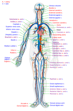
The human circulatory system (simplified). Red indicates oxygenated blood carried inarteries, blue indicates deoxygenated blood carried in veins. Capillaries, which join the arteries and veins, and the lymphatic vesselsare not shown.
| |
| Details | |
| Latin | Systema cardiovasculare |
| Identifiers | |
| Anatomical terminology | |
The circulatory system, also called the cardiovascular system, is an organ system that permits blood to circulate and transport nutrients (such as amino acids and electrolytes), oxygen, carbon dioxide, hormones, and blood cells to and from the cells in the body to provide nourishment and help in fighting diseases, stabilize temperature and pH, and maintain homeostasis. The study of the blood flow is called hemodynamics. The study of the properties of the blood flow is called hemorheology.
The circulatory system is often seen to comprise both the cardiovascular system, which distributes blood, and the lymphatic system, which circulateslymph.[1] These are two separate systems. The passage of lymph for example takes a lot longer than that of blood.[2] Blood is a fluid consisting of plasma, red blood cells, white blood cells, and platelets that is circulated by the heartthrough the vertebrate vascular system, carrying oxygen and nutrients to and waste materials away from all body tissues. Lymph is essentially recycled excess blood plasma after it has been filtered from the interstitial fluid (between cells) and returned to the lymphatic system. The cardiovascular (from Latin words meaning 'heart' and 'vessel') system comprises the blood, heart, andblood vessels.[3] The lymph, lymph nodes, and lymph vessels form the lymphatic system, which returns filtered blood plasma from the interstitial fluid (between cells) as lymph.
While humans, as well as other vertebrates, have a closed cardiovascular system (meaning that the blood never leaves the network of arteries, veins andcapillaries), some invertebrate groups have an open cardiovascular system. The lymphatic system, on the other hand, is an open system providing an accessory route for excess interstitial fluid to get returned to the blood.[4] The more primitive, diploblastic animal phyla lack circulatory systems.
Contents
Structure
Cardiovascular system
The essential components of the human cardiovascular system are the heart,blood and blood vessels.[5] It includes thepulmonary circulation, a "loop" through the lungs where blood is oxygenated; and the systemic circulation, a "loop" through the rest of the body to provideoxygenated blood. The systemic circulation can also be seen to function in two parts–a macrocirculation and amicrocirculation. An average adult contains five to six quarts (roughly 4.7 to 5.7 liters) of blood, accounting for approximately 7% of their total body weight.[6] Blood consists of plasma, red blood cells, white blood cells, and platelets. Also, the digestive system works with the circulatory system to provide the nutrients the system needs to keep the heart pumping.[7]
The cardiovascular systems of humans are closed, meaning that the blood never leaves the network of blood vessels. In contrast, oxygen and nutrients diffuse across the blood vessel layers and enter interstitial fluid, which carries oxygen and nutrients to the target cells, and carbon dioxide and wastes in the opposite direction. The other component of the circulatory system, the lymphatic system, is open.
Arteries
See also: Arterial tree
Oxygenated blood enters the systemic circulation when leaving the left ventricle, through the aortic semilunar valve. The first part of the systemic circulation is the aorta, a massive and thick-walled artery. The aorta arches and branches into major arteries to the upper body before passing through the diaphragm, where it branches further into arteries which supply the lower parts of the body.
Capillaries
Arteries branch into small passages called arterioles and then into the capillaries.[8] The capillaries merge to bring blood into the venous system.[9]
Veins
After their passage through body tissues, capillaries merge once again into venules, which continue to merge into veins. The venous system finally coalesces into two major veins: the superior vena cava (roughly speaking draining the areas above the heart) and the inferior vena cava (roughly speaking from areas below the heart). These two great vessels empty into the right atrium of the heart.
Coronary vessels
Main article: Coronary circulation
The heart itself is supplied with oxygen and nutrients through a small "loop" of the systemic circulation.
Portal veins
The general rule is that arteries from the heart branch out into capillaries, which collect into veins leading back to the heart.Portal veins are a slight exception to this. In humans the only significant example is the hepatic portal vein which combines from capillaries around the gut where the blood absorbs the various products of digestion; rather than leading directly back to the heart, the hepatic portal vein branches into a second capillary system in the liver.
Heart
Main article: Heart
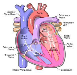
The heart pumps oxygenated blood to the body and deoxygenated blood to the lungs. In the human heart there is one atrium and one ventricle for each circulation, and with both a systemic and a pulmonary circulation there are four chambers in total: left atrium, left ventricle, right atrium and right ventricle. The right atrium is the upper chamber of the right side of the heart. The blood that is returned to the right atrium is deoxygenated (poor in oxygen) and passed into the right ventricle to be pumped through the pulmonary artery to the lungs for re-oxygenation and removal of carbon dioxide. The left atrium receives newly oxygenated blood from the lungs as well as the pulmonary vein which is passed into the strong left ventricle to be pumped through the aorta to the different organs of the body.
The coronary circulation system provides a blood supply to the heart muscleitself. The coronary circulation begins near the origin of the aorta by two arteries: the right coronary artery and the left coronary artery. After nourishing the heart muscle, blood returns through the coronary veins into the coronary sinus and from this one into the right atrium. Back flow of blood through its opening during atrial systole is prevented by the Thebesian valve. The smallest cardiac veinsdrain directly into the heart chambers.[7]
Pulmonary circulation
Main article: Pulmonary circulation
The circulatory system of the lungs is the portion of the cardiovascular system in which oxygen-depleted blood is pumped away from the heart, via the pulmonary artery, to the lungs and returned, oxygenated, to the heart via thepulmonary vein.
Oxygen deprived blood from the superior and inferior vena cava enters the right atrium of the heart and flows through the tricuspid valve (right atrioventricular valve) into the right ventricle, from which it is then pumped through thepulmonary semilunar valve into the pulmonary artery to the lungs. Gas exchange occurs in the lungs, whereby CO2 is released from the blood, and oxygen is absorbed. The pulmonary vein returns the now oxygen-rich blood to the left atrium.[7]
A separate system known as the bronchial circulation supplies blood to the tissue of the larger airways of the lung.
Systemic circulation
The systemic circulation is the circulation of the blood to all parts of the body except the lungs. Systemic circulation is the portion of the cardiovascular system which transports oxygenated blood away from the heart through the aorta from the left ventricle where the blood has been previously deposited from pulmonary circulation, to the rest of the body, and returns oxygen-depleted blood back to the heart.[7]
Brain
Main article: Cerebral circulation
The brain has a dual blood supply that comes from arteries at its front and back. These are called the "anterior" and "posterior" circulation respectively. The anterior circulation arises from the internal carotid arteries and supplies the front of the brain. The posterior circulation arises from the vertebral arteries, and supplies the back of the brain and brainstem. The circulation from the front and the back join together (anastomise) at the Circle of Willis.
Kidneys
The renal circulation receives around 20% of the cardiac output. It branches from the abdominal aorta and returns blood to the ascending vena cava. It is the blood supply to the kidneys, and contains many specialized blood vessels.
Lymphatic system
The lymphatic system is part of the circulatory system. It is a network of lymphatic vessels and lymph capillaries, lymph nodes and organs, and lymphatic tissues and circulating lymph. One of its major functions is to carry the lymph, draining and returning interstitial fluid back towards the heart for return to the cardiovascular system, by emptying into the lymphatic ducts. Its other main function is in the immune system.
Physiology
Main article: Blood § Oxygen transport
About 98.5% of the oxygen in a sample of arterial blood in a healthy human, breathing air at sea-level pressure, is chemically combined with hemoglobinmolecules. About 1.5% is physically dissolved in the other blood liquids and not connected to hemoglobin. The hemoglobin molecule is the primary transporter of oxygen in mammals and many other species.
Development
Main article: Fetal circulation
The development of the circulatory system starts with vasculogenesis in the embryo. The human arterial and venous systems develop from different areas in the embryo. The arterial system develops mainly from the aortic arches, six pairs of arches which develop on the upper part of the embryo. The venous system arises from three bilateral veins during weeks 4 – 8 of embryogenesis. Fetal circulation begins within the 8th week of development. Fetal circulation does not include the lungs, which are bypassed via the truncus arteriosus. Before birth the fetus obtains oxygen (and nutrients) from the mother through theplacenta and the umbilical cord.[10]
Arterial development
Main article: Aortic arches
The human arterial system originates from the aortic arches and from the dorsal aortae starting from week 4 of embryonic life. The first and second aortic arches regress and forms only the maxillary arteries and stapedial arteries respectively. The arterial system itself arises from aortic arches 3, 4 and 6 (aortic arch 5 completely regresses).
The dorsal aortae, present on the dorsal side of the embryo, are initially present on both sides of the embryo. They later fuse to form the basis for the aorta itself. Approximately thirty smaller arteries branch from this at the back and sides. These branches form the intercostal arteries, arteries of the arms and legs, lumbar arteries and the lateral sacral arteries. Branches to the sides of the aorta will form the definitive renal, suprarenal and gonadal arteries. Finally, branches at the front of the aorta consist of the vitelline arteries and umbilical arteries. The vitelline arteries form the celiac, superior andinferior mesenteric arteries of the gastrointestinal tract. After birth, the umbilical arteries will form the internal iliac arteries.
Venous development
The human venous system develops mainly from the vitelline veins, the umbilical veins and the cardinal veins, all of which empty into the sinus venosus.
Clinical significance
Many diseases affect the circulatory system. This includes cardiovascular disease, affecting the cardiovascular system, andlymphatic disease affecting the lymphatic system. Cardiologists are medical professionals which specialise in the heart, andcardiothoracic surgeons specialise in operating on the heart and its surrounding areas. Vascular surgeons focus on other parts of the circulatory system.
Cardiovascular disease
Main article: Cardiovascular disease
Diseases affecting the cardiovascular system are called cardiovascular disease.
Many of these diseases are called "lifestyle diseases" because they develop over time and are related to a person's exercise habits, diet, whether they smoke, and other lifestyle choices a person makes. Atherosclerosis is the precursor to many of these diseases. It is where small atheromatous plaques build up in the walls of medium and large arteries. This may eventually grow or rupture to occlude the arteries. It is also a risk factor for acute coronary syndromes, which are diseases which are characterised by a sudden deficit of oxygenated blood to the heart tissue. Atherosclerosis is also associated with problems such as aneurysm formation or splitting ("dissection") of arteries.
Another major cardiovascular disease involves the creation of a clot, called a "thrombus". These can originate in veins or arteries. Deep venous thrombosis, which mostly occurs in the legs, is one cause of clots in the veins of the legs, particularly when a person has been stationary for a long time. These clots may embolise, meaning travel to another location in the body. The results of this may include pulmonary embolus, transient ischaemic attacks, or stroke.
Cardiovascular diseases may also be congenital in nature, such as heart defects or persistent fetal circulation, where the circulatory changes that are supposed to happen after birth do not. Not all congenital changes to the circulatory system are associated with diseases, a large number are anatomical variations.
Measurement techniques
The function and health of the circulatory system and its parts are measured in a variety of manual and automated ways. These include simple methods such as those that are part of the cardiovascular examination, including the taking of a person's pulse as an indicator of a person's heart rate, the taking of blood pressurethrough a sphygmomanometer or the use of a stethoscope to listen to the heart formurmurs which may indicate problems with the heart's valves. An electrocardiogramcan also be used to evaluate the way in which electricity is conducted through the heart.
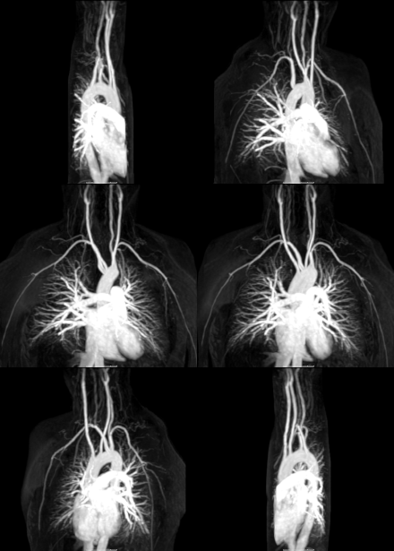
Other more invasive means can also be used. A cannula or catheter inserted into an artery may be used to measure pulse pressure or pulmonary wedge pressures. Angiography, which involves injecting a dye into an artery to visualise an arterial tree, can be used in the heart (coronary angiography) or brain. At the same time as the arteries are visualised, blockages or narrowings may be fixed through the insertion of stents, and active bleeds may be managed by the insertion of coils. An MRI may be used to image arteries, called an MRI angiogram. For evaluation of the blood supply to the lungs a CT pulmonary angiogram may be used.
Ultrasound can also be used, particularly to identify the health of blood vessels, and a Doppler ultrasound of the carotid arteries or Doppler ultrasound of the lower limbs can be used to evaluate for narrowing of the carotid arteries or thrombusformation in the legs, respectively.
Surgery
There are a number of surgical procedures performed on the circulatory system:
- Coronary artery bypass surgery
- Coronary stent used in angioplasty
- Vascular surgery
- Vein stripping
- Cosmetic procedures
Cardiovascular procedures are more likely to performed in the inpatient setting than in an ambulatory care setting; in the United States, only 28% of cardiovascular surgeries were performed in the ambulatory care setting.[11]
Society and culture
| This section requires expansion.(March 2015) |
A number of alternative medical systems such as Chinese medicine view the circulatory system in different ways.
Other animals
Other vertebrates
The circulatory systems of all vertebrates, as well as of annelids (for example,earthworms) and cephalopods (squids, octopuses and relatives) are closed, just as in humans. Still, the systems of fish, amphibians, reptiles, and birds show various stages of the evolution of the circulatory system.
In fish, the system has only one circuit, with the blood being pumped through the capillaries of the gills and on to the capillaries of the body tissues. This is known assingle cycle circulation. The heart of fish is, therefore, only a single pump (consisting of two chambers).
In amphibians and most reptiles, a double circulatory system is used, but the heart is not always completely separated into two pumps. Amphibians have a three-chambered heart.
In reptiles, the ventricular septum of the heart is incomplete and the pulmonary artery is equipped with a sphincter muscle. This allows a second possible route of blood flow. Instead of blood flowing through the pulmonary artery to the lungs, the sphincter may be contracted to divert this blood flow through the incomplete ventricular septum into the left ventricle and out through the aorta. This means the blood flows from the capillaries to the heart and back to the capillaries instead of to the lungs. This process is useful to ectothermic (cold-blooded) animals in the regulation of their body temperature.
Birds and mammals show complete separation of the heart into two pumps, for a total of four heart chambers; it is thought[citation needed] that the four-chambered heart of birds evolved independently from that of mammals.
Open circulatory system
See also: Hemolymph
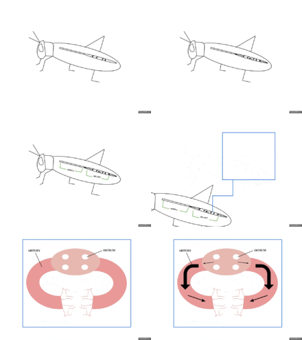
The open circulatory system is a system in which a fluid in a cavity called the hemocoel bathes the organs directly with oxygen and nutrients and there is no distinction between blood and interstitial fluid; this combined fluid is called hemolymphor haemolymph.[12] Muscular movements by the animal during locomotion can facilitate hemolymph movement, but diverting flow from one area to another is limited. When the heart relaxes, blood is drawn back toward the heart through open-ended pores (ostia).
Hemolymph fills all of the interior hemocoel of the body and surrounds all cells. Hemolymph is composed of water, inorganicsalts (mostly Na+, Cl−, K+, Mg2+, and Ca2+), and organic compounds (mostly carbohydrates, proteins, and lipids). The primary oxygen transporter molecule is hemocyanin.
There are free-floating cells, the hemocytes, within the hemolymph. They play a role in the arthropod immune system.
Absence of circulatory system[edit]
Circulatory systems are absent in some animals, including flatworms (phylumPlatyhelminthes). Their body cavity has no lining or enclosed fluid. Instead a muscularpharynx leads to an extensively brancheddigestive system that facilitates directdiffusion of nutrients to all cells. The flatworm's dorso-ventrally flattened body shape also restricts the distance of any cell from the digestive system or the exterior of the organism. Oxygen can diffuse from the surrounding water into the cells, andcarbon dioxide can diffuse out. Consequently every cell is able to obtain nutrients, water and oxygen without the need of a transport system.
Some animals, such as jellyfish, have more extensive branching from theirgastrovascular cavity (which functions as both a place of digestion and a form of circulation), this branching allows for bodily fluids to reach the outer layers, since the digestion begins in the inner layers.
History
The earliest known writings on the circulatory system are found in the Ebers Papyrus(16th century BCE), an ancient Egyptian medical papyrus containing over 700 prescriptions and remedies, both physical and spiritual. In the papyrus, it acknowledges the connection of the heart to the arteries. The Egyptians thought air came in through the mouth and into the lungs and heart. From the heart, the air travelled to every member through the arteries. Although this concept of the circulatory system is only partially correct, it represents one of the earliest accounts of scientific thought.
In the 6th century BCE, the knowledge of circulation of vital fluids through the body was known to the Ayurvedic physician Sushruta in ancient India.[13] He also seems to have possessed knowledge of the arteries, described as 'channels' by Dwivedi & Dwivedi (2007).[13] The valves of the heart were discovered by a physician of theHippocratean school around the 4th century BCE. However their function was not properly understood then. Because blood pools in the veins after death, arteries look empty. Ancient anatomists assumed they were filled with air and that they were for transport of air.
The Greek physician, Herophilus, distinguished veins from arteries but thought that the pulse was a property of arteries themselves. Greek anatomist Erasistratusobserved that arteries that were cut during life bleed. He ascribed the fact to the phenomenon that air escaping from an artery is replaced with blood that entered by very small vessels between veins and arteries. Thus he apparently postulated capillaries but with reversed flow of blood.[14]
In 2nd century AD Rome, the Greek physician Galen knew that blood vessels carried blood and identified venous (dark red) and arterial (brighter and thinner) blood, each with distinct and separate functions. Growth and energy were derived from venous blood created in the liver from chyle, while arterial blood gave vitality by containing pneuma (air) and originated in the heart. Blood flowed from both creating organs to all parts of the body where it was consumed and there was no return of blood to the heart or liver. The heart did not pump blood around, the heart's motion sucked blood in during diastole and the blood moved by the pulsation of the arteries themselves.
Galen believed that the arterial blood was created by venous blood passing from the left ventricle to the right by passing through 'pores' in the interventricular septum, air passed from the lungs via the pulmonary artery to the left side of the heart. As the arterial blood was created 'sooty' vapors were created and passed to the lungs also via the pulmonary artery to be exhaled.
In 1025, The Canon of Medicine by the Persian physician, Avicenna, "erroneously accepted the Greek notion regarding the existence of a hole in the ventricular septum by which the blood traveled between the ventricles." Despite this, Avicenna "correctly wrote on the cardiac cycles and valvular function", and "had a vision of blood circulation" in his Treatise on Pulse.[15][verification needed] While also refining Galen's erroneous theory of the pulse, Avicenna provided the first correct explanation of pulsation: "Every beat of the pulse comprises two movements and two pauses. Thus, expansion : pause : contraction : pause. [...] The pulse is a movement in the heart and arteries ... which takes the form of alternate expansion and contraction."[16]
In 1242, the Arabian physician, Ibn al-Nafis, became the first person to accurately describe the process of pulmonary circulation, for which he is sometimes considered the father of circulatory physiology.[17][not in citation given] Ibn al-Nafis stated in his Commentary on Anatomy in Avicenna's Canon:
In addition, Ibn al-Nafis had an insight into what would become a larger theory of the capillary circulation. He stated that "there must be small communications or pores (manafidh in Arabic) between the pulmonary artery and vein," a prediction that preceded the discovery of the capillary system by more than 400 years.[18] Ibn al-Nafis' theory, however, was confined to blood transit in the lungs and did not extend to the entire body.
Michael Servetus was the first European to describe the function of pulmonary circulation, although his achievement was not widely recognized at the time, for a few reasons. He firstly described it in the "Manuscript of Paris"[19][20] (near 1546), but this work was never published. And later he published this description, but in a theological treatise, Christianismi Restitutio, not in a book on medicine. Only three copies of the book survived, the rest were burned shortly after its publication in 1553 because of persecution of Servetus by religious authorities. Better known was its discovery by Vesalius's successor atPadua, Realdo Colombo, in 1559.
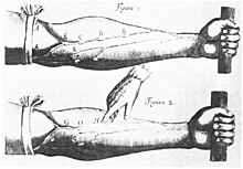
Finally, William Harvey, a pupil of Hieronymus Fabricius (who had earlier described the valves of the veins without recognizing their function), performed a sequence of experiments, and published Exercitatio Anatomica de Motu Cordis et Sanguinis in Animalibus in 1628, which "demonstrated that there had to be a direct connection between the venous and arterial systems throughout the body, and not just the lungs. Most importantly, he argued that the beat of the heart produced a continuous circulation of blood through minute connections at the extremities of the body. This is a conceptual leap that was quite different from Ibn al-Nafis' refinement of the anatomy and bloodflow in the heart and lungs."[21] This work, with its essentially correct exposition, slowly convinced the medical world. However, Harvey was not able to identify the capillary system connecting arteries and veins; these were later discovered by Marcello Malpighi in 1661.
In 1956, André Frédéric Cournand, Werner Forssmann and Dickinson W. Richards were awarded the Nobel Prize in Medicine "for their discoveries concerning heart catheterization and pathological changes in the circulatory system."[22]
Blood
For other uses, see Blood (disambiguation).
| Blood | |
|---|---|
 | |
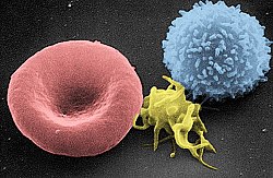
A scanning electron microscope (SEM) image of a normal red blood cell, a platelet, and awhite blood cell.
| |
| Details | |
| Latin | haema |
| Identifiers | |
| Anatomical terminology | |
Blood is a bodily fluid in animals that delivers necessary substances such asnutrients and oxygen to the cells and transports metabolic waste products away from those same cells.[1] When it reaches the lungs, gas exchange occurs when carbon dioxide is diffused out of the blood into the pulmonary alveoli and oxygen is diffused into the blood. This oxygenated blood is pumped to the left hand side of the heart in the pulmonary vein and enters the left atrium. From here it passes through the mitral valve, through the ventricle and taken all around the body by the aorta. Blood contains antibodies, nutrients, oxygen and much more to help the body work.[citation needed]
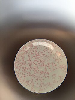
In vertebrates, it is composed of blood cells suspended in blood plasma. Plasma, which constitutes 55% of blood fluid, is mostly water (92% by volume),[2] and contains dissipated proteins, glucose, mineral ions, hormones, carbon dioxide (plasma being the main medium for excretory product transportation), and blood cells themselves. Albumin is the main protein in plasma, and it functions to regulate the colloidal osmotic pressure of blood. The blood cells are mainly red blood cells (also called RBCs or erythrocytes),white blood cells (also called WBCs or leukocytes) and platelets. The most abundant cells in vertebrate blood are red blood cells. These containhemoglobin, an iron-containing protein, which facilitates oxygen transport by reversibly binding to this respiratory gas and greatly increasing its solubility in blood. In contrast, carbon dioxide is almost entirely transported extracellularly dissolved in plasma as bicarbonate ion.[citation needed]
Vertebrate blood is bright red when its haemoglobin is oxygenated and dark red when it is deoxygenated. Some animals, such as crustaceans andmollusks, use hemocyanin to carry oxygen, instead of hemoglobin. Insects and some mollusks use a fluid called hemolymph instead of blood, the difference being that hemolymph is not contained in a closed circulatory system. In most insects, this "blood" does not contain oxygen-carrying molecules such as hemoglobin because their bodies are small enough for their tracheal system to suffice for supplying oxygen.
Jawed vertebrates have an adaptive immune system, based largely on white blood cells. White blood cells help to resist infections and parasites. Platelets are important in the clotting of blood. Arthropods, using hemolymph, have hemocytes as part of their immune system.
Blood is circulated around the body through blood vessels by the pumping action of the heart. In animals with lungs, arterialblood carries oxygen from inhaled air to the tissues of the body, and venous blood carries carbon dioxide, a waste product of metabolism produced by cells, from the tissues to the lungs to be exhaled.
Medical terms related to blood often begin with hemo- or hemato- (also spelled haemo- and haemato-) from the Greek word αἷμα (haima) for "blood". In terms of anatomy and histology, blood is considered a specialized form of connective tissue, given its origin in the bones and the presence of potential molecular fibers in the form of fibrinogen.
Contents
Functions
Blood performs many important functions within the body including:
- Supply of oxygen to tissues (bound to hemoglobin, which is carried in red cells)
- Supply of nutrients such as glucose, amino acids, and fatty acids (dissolved in the blood or bound to plasma proteins (e.g., blood lipids))
- Removal of waste such as carbon dioxide, urea, and lactic acid
- Immunological functions, including circulation of white blood cells, and detection of foreign material by antibodies
- Coagulation, the response to a broken blood vessel, the conversion of blood from a liquid to a semi-solid gel to stop bleeding.
- Messenger functions, including the transport of hormones and the signaling oftissue damage
- Regulation of body pH
- Regulation of core body temperature
- Hydraulic functions
Constituents
See also: Reference ranges for common blood tests
Blood accounts for 7% of the human body weight,[3][4] with an average density of approximately 1060 kg/m3, very close to pure water's density of 1000 kg/m3.[5] The average adult has a blood volume of roughly 5 litres (11 US pt),[4] which is composed of plasma and several kinds of cells. These blood cells (which are also called corpusclesor "formed elements") consist of erythrocytes (red blood cells, RBCs), leukocytes (white blood cells), and thrombocytes (platelets). By volume, the red blood cells constitute about 45% of whole blood, the plasma about 54.3%, and white cells about 0.7%.
Whole blood (plasma and cells) exhibits non-Newtonian fluid dynamics; its flow properties are adapted to flow effectively through tiny capillary blood vessels with less resistance than plasma by itself. In addition, if all human hemoglobin were free in the plasma rather than being contained in RBCs, the circulatory fluid would be too viscous for the cardiovascular system to function effectively.
Cells
Further information: Complete blood count
One microliter of blood contains:
- 4.7 to 6.1 million (male), 4.2 to 5.4 million (female) erythrocytes:[6] Red blood cells contain the blood's hemoglobinand distribute oxygen. Mature red blood cells lack a nucleus and organelles in mammals. The red blood cells (together with endothelial vessel cells and other cells) are also marked by glycoproteins that define the different blood types. The proportion of blood occupied by red blood cells is referred to as the hematocrit, and is normally about 45%. The combined surface area of all red blood cells of the human body would be roughly 2,000 times as great as the body's exterior surface.[7]
- 4,000–11,000 leukocytes:[8] White blood cells are part of the body's immune system; they destroy and remove old or aberrant cells and cellular debris, as well as attack infectious agents (pathogens) and foreign substances. The cancer of leukocytes is called leukemia.
- 200,000–500,000 thrombocytes:[8] Also called platelets, they take part in blood clotting (coagulation). Fibrin from the coagulation cascade creates a mesh over the platelet plug.
| Parameter | Value |
|---|---|
| Hematocrit |
45 ± 7 (38–52%) for males
42 ± 5 (37–47%) for females |
| pH | 7.35–7.45 |
| base excess | −3 to +3 |
| PO2 | 10–13 kPa (80–100 mm Hg) |
| PCO2 | 4.8–5.8 kPa (35–45 mm Hg) |
| HCO3− | 21–27 mM |
| Oxygen saturation |
Oxygenated: 98–99%
Deoxygenated: 75% |
Plasma
Main article: Blood plasma
About 55% of blood is blood plasma, a fluid that is the blood's liquid medium, which by itself is straw-yellow in color. The blood plasma volume totals of 2.7–3.0 liters (2.8–3.2 quarts) in an average human. It is essentially an aqueous solution containing 92% water, 8% blood plasmaproteins, and trace amounts of other materials. Plasma circulates dissolved nutrients, such as glucose, amino acids, and fatty acids(dissolved in the blood or bound to plasma proteins), and removes waste products, such as carbon dioxide, urea, and lactic acid.
Other important components include:
- Serum albumin
- Blood-clotting factors (to facilitate coagulation)
- Immunoglobulins (antibodies)
- lipoprotein particles
- Various other proteins
- Various electrolytes (mainly sodium and chloride)
The term serum refers to plasma from which the clotting proteins have been removed. Most of the proteins remaining are albumin and immunoglobulins.
pH values
See also: Acid-base homeostasis
Blood pH is regulated to stay within the narrow range of 7.35 to 7.45, making it slightly basic.[9][10] Blood that has a pH below 7.35 is too acidic, whereas blood pH above 7.45 is too basic. Blood pH, partial pressure of oxygen (pO2), partial pressure of carbon dioxide (pCO2), and HCO3− are carefully regulated by a number of homeostatic mechanisms, which exert their influence principally through the respiratory system and the urinary system in order to control the acid-base balance and respiration. An arterial blood gas test will measure these. Plasma also circulates hormones transmitting their messages to various tissues. The list of normal reference ranges for various blood electrolytes is extensive.
Blood in non-mammalian vertebrates
Human blood is typical of that of mammals, although the precise details concerning cell numbers, size, protein structure, and so on, vary somewhat between species. In non-mammalian vertebrates, however, there are some key differences:[11]
- Red blood cells of non-mammalian vertebrates are flattened and ovoid in form, and retain their cell nuclei
- There is considerable variation in the types and proportions of white blood cells; for example, acidophils are generally more common than in humans
- Platelets are unique to mammals; in other vertebrates, small nucleated, spindle cells called thrombocytes are responsible for blood clotting instead
Physiology[edit]
Cardiovascular system
Main article: Circulatory system
Blood is circulated around the body through blood vessels by the pumping action of the heart. In humans, blood is pumped from the strong left ventricle of the heart through arteries to peripheral tissues and returns to the right atrium of the heartthrough veins. It then enters the right ventricle and is pumped through thepulmonary artery to the lungs and returns to the left atrium through the pulmonary veins. Blood then enters the left ventricle to be circulated again. Arterial blood carries oxygen from inhaled air to all of the cells of the body, and venous bloodcarries carbon dioxide, a waste product of metabolism by cells, to the lungs to be exhaled. However, one exception includes pulmonary arteries, which contain the most deoxygenated blood in the body, while the pulmonary veins contain oxygenated blood.
Additional return flow may be generated by the movement of skeletal muscles, which can compress veins and push blood through the valves in veins toward the right atrium.
The blood circulation was famously described by William Harvey in 1628.[12]
Production and degradation of blood cells
In vertebrates, the various cells of blood are made in the bone marrow in a process called hematopoiesis, which includes erythropoiesis, the production of red blood cells; and myelopoiesis, the production of white blood cells and platelets. During childhood, almost every human bone produces red blood cells; as adults, red blood cell production is limited to the larger bones: the bodies of the vertebrae, the breastbone (sternum), the ribcage, the pelvic bones, and the bones of the upper arms and legs. In addition, during childhood, the thymus gland, found in themediastinum, is an important source of T lymphocytes.[13] The proteinaceous component of blood (including clotting proteins) is produced predominantly by theliver, while hormones are produced by the endocrine glands and the watery fraction is regulated by the hypothalamus and maintained by the kidney.
Healthy erythrocytes have a plasma life of about 120 days before they are degraded by the spleen, and the Kupffer cells in the liver. The liver also clears some proteins, lipids, and amino acids. The kidney actively secretes waste products into the urine.
Oxygen transport
About 98.5% of the oxygen in a sample of arterial blood in a healthy human breathing air at sea-level pressure is chemically combined with the Hgb. About 1.5% is physically dissolved in the other blood liquids and not connected to Hgb. Thehemoglobin molecule is the primary transporter of oxygen in mammals and many other species (for exceptions, see below). Hemoglobin has an oxygen binding capacity of between 1.36 and 1.37 ml O2 per gram hemoglobin,[14] which increases the total blood oxygen capacity seventyfold,[15] compared to if oxygen solely were carried by its solubility of 0.03 ml O2 per liter blood per mm Hg partial pressure of oxygen (approximately 100 mm Hg in arteries).[15]
With the exception of pulmonary and umbilical arteries and their corresponding veins, arteries carry oxygenated blood away from the heart and deliver it to the body via arterioles and capillaries, where the oxygen is consumed; afterwards, venules, and veins carry deoxygenated blood back to the heart.
Under normal conditions in adult humans at rest; hemoglobin in blood leaving the lungs is about 98–99% saturated with oxygen, achieving an oxygen delivery of between 950 and 1150 ml/min[16] to the body. In a healthy adult at rest, oxygen consumption is approximately 200 - 250 ml/min,[16] and deoxygenated blood returning to the lungs is still approximately 75%[17][18] (70 to 78%)[16] saturated. Increased oxygen consumption during sustained exercise reduces the oxygen saturation of venous blood, which can reach less than 15% in a trained athlete; although breathing rate and blood flow increase to compensate, oxygen saturation in arterial blood can drop to 95% or less under these conditions.[19] Oxygen saturation this low is considered dangerous in an individual at rest (for instance, during surgery under anesthesia). Sustained hypoxia (oxygenation of less than 90%), is dangerous to health, and severe hypoxia (saturations of less than 30%) may be rapidly fatal.[20]
A fetus, receiving oxygen via the placenta, is exposed to much lower oxygen pressures (about 21% of the level found in an adult's lungs), and, so, fetuses produce another form of hemoglobin with a much higher affinity for oxygen (hemoglobin F) in order to function under these conditions.[21]
Carbon dioxide transport
CO2 is carried in blood in three different ways. (The exact percentages vary depending whether it is arterial or venous blood). Most of it (about 70%) is converted to bicarbonate ions HCO−
3 by the enzyme carbonic anhydrase in the red blood cells by the reaction CO2 + H2O → H2CO3 → H+ + HCO−
3; about 7% is dissolved in the plasma; and about 23% is bound to hemoglobin as carbamino compounds.[22] Hemoglobin, the main oxygen-carrying molecule in red blood cells, carries both oxygen and carbon dioxide. However, the CO2 bound to hemoglobin does not bind to the same site as oxygen. Instead, it combines with the N-terminal groups on the four globin chains. However, because of allosteric effects on the hemoglobin molecule, the binding of CO2 decreases the amount of oxygen that is bound for a given partial pressure of oxygen. The decreased binding to carbon dioxide in the blood due to increased oxygen levels is known as the Haldane effect, and is important in the transport of carbon dioxide from the tissues to the lungs. A rise in the partial pressure of CO2or a lower pH will cause offloading of oxygen from hemoglobin, which is known as the Bohr effect.
Transport of hydrogen ions
Some oxyhemoglobin loses oxygen and becomes deoxyhemoglobin. Deoxyhemoglobin binds most of the hydrogen ions as it has a much greater affinity for more hydrogen than does oxyhemoglobin.
Lymphatic system
Main article: Lymphatic system
In mammals, blood is in equilibrium with lymph, which is continuously formed in tissues from blood by capillary ultrafiltration. Lymph is collected by a system of small lymphatic vessels and directed to the thoracic duct, which drains into the leftsubclavian vein where lymph rejoins the systemic blood circulation.
Thermoregulation
Blood circulation transports heat throughout the body, and adjustments to this flow are an important part ofthermoregulation. Increasing blood flow to the surface (e.g., during warm weather or strenuous exercise) causes warmer skin, resulting in faster heat loss. In contrast, when the external temperature is low, blood flow to the extremities and surface of the skin is reduced and to prevent heat loss and is circulated to the important organs of the body, preferentially.
Hydraulic functions
The restriction of blood flow can also be used in specialized tissues to cause engorgement, resulting in an erection of that tissue; examples are the erectile tissue in the penis and clitoris.
Another example of a hydraulic function is the jumping spider, in which blood forced into the legs under pressure causes them to straighten for a powerful jump, without the need for bulky muscular legs.[23]
Invertebrates[edit]
In insects, the blood (more properly called hemolymph) is not involved in the transport of oxygen. (Openings called tracheaeallow oxygen from the air to diffuse directly to the tissues). Insect blood moves nutrients to the tissues and removes waste products in an open system.
Other invertebrates use respiratory proteins to increase the oxygen-carrying capacity. Hemoglobin is the most common respiratory protein found in nature. Hemocyanin (blue) contains copper and is found in crustaceans and mollusks. It is thought that tunicates (sea squirts) might use vanabins (proteins containing vanadium) for respiratory pigment (bright-green, blue, or orange).
In many invertebrates, these oxygen-carrying proteins are freely soluble in the blood; in vertebrates they are contained in specialized red blood cells, allowing for a higher concentration of respiratory pigments without increasing viscosity or damaging blood filtering organs like the kidneys.
Giant tube worms have unusual hemoglobins that allow them to live in extraordinary environments. These hemoglobins also carry sulfides normally fatal in other animals.
Color[edit]
The coloring matter of blood (hemochrome) is largely due to the protein in the blood responsible for oxygen transport. Different groups of organisms use different proteins.
Hemoglobin[edit]
Main article: Hemoglobin
Hemoglobin is the principal determinant of the color of blood in vertebrates. Each molecule has four heme groups, and their interaction with various molecules alters the exact color. In vertebrates and other hemoglobin-using creatures, arterial blood and capillary blood are bright red, as oxygen imparts a strong red color to the heme group. Deoxygenated blood is a darker shade of red; this is present in veins, and can be seen during blood donation and when venous blood samples are taken. This is because the spectrum of light absorbed by hemoglobin differs between the oxygenated and deoxygenated states.[24]
Blood in carbon monoxide poisoning is bright red, because carbon monoxide causes the formation of carboxyhemoglobin. In cyanide poisoning, the body cannot utilize oxygen, so the venous blood remains oxygenated, increasing the redness. There are some conditions affecting the heme groups present in hemoglobin that can make the skin appear blue—a symptom called cyanosis. If the heme is oxidized,methaemoglobin, which is more brownish and cannot transport oxygen, is formed. In the rare condition sulfhemoglobinemia, arterial hemoglobin is partially oxygenated, and appears dark red with a bluish hue.
Veins close to the surface of the skin appear blue for a variety of reasons. However, the factors that contribute to this alteration of color perception are related to the light-scattering properties of the skin and the processing of visual input by the visual cortex, rather than the actual color of the venous blood.[25]
Skinks in the genus Prasinohaema have green blood due to a buildup of the waste product biliverdin.[26]
Hemocyanin[edit]
Main article: Hemocyanin
The blood of most mollusks – including cephalopods and gastropods – as well as some arthropods, such as horseshoe crabs, is blue, as it contains the copper-containing protein hemocyanin at concentrations of about 50 grams per liter.[27]Hemocyanin is colorless when deoxygenated and dark blue when oxygenated. The blood in the circulation of these creatures, which generally live in cold environments with low oxygen tensions, is grey-white to pale yellow,[27] and it turns dark blue when exposed to the oxygen in the air, as seen when they bleed.[27] This is due to change in color of hemocyanin when it is oxidized.[27] Hemocyanin carries oxygen in extracellular fluid, which is in contrast to the intracellular oxygen transport in mammals by hemoglobin in RBCs.[27]
Chlorocruorin[edit]
Main article: Chlorocruorin
The blood of most annelid worms and some marine polychaetes use chlorocruorin to transport oxygen. It is green in color in dilute solutions.[28]
Hemerythrin[edit]
Main article: Hemerythrin
Hemerythrin is used for oxygen transport in the marine invertebrates sipunculids, priapulids, brachiopods, and the annelid worm, magelona. Hemerythrin is violet-pink when oxygenated.[28]
Hemovanadin[edit]
Main article: Hemovanadin
The blood of some species of ascidians and tunicates, also known as sea squirts, contains proteins called vanabins. These proteins are based on vanadium, and give the creatures a concentration of vanadium in their bodies 100 times higher than the surrounding sea water. Unlike hemocyanin and hemoglobin, hemovanadin is not an oxygen carrier. When exposed to oxygen, however, vanabins turn a mustard yellow.
Pathology[edit]
General medical disorders[edit]
- Disorders of volume
- Injury can cause blood loss through bleeding.[29] A healthy adult can lose almost 20% of blood volume (1 L) before the first symptom, restlessness, begins, and 40% of volume (2 L) before shock sets in. Thrombocytes are important for blood coagulation and the formation of blood clots, which can stop bleeding. Trauma to the internal organs or bones can cause internal bleeding, which can sometimes be severe.
- Dehydration can reduce the blood volume by reducing the water content of the blood. This would rarely result inshock (apart from the very severe cases) but may result in orthostatic hypotension and fainting.
- Disorders of circulation
- Shock is the ineffective perfusion of tissues, and can be caused by a variety of conditions including blood loss, infection, poor cardiac output.
- Atherosclerosis reduces the flow of blood through arteries, because atheroma lines arteries and narrows them. Atheroma tends to increase with age, and its progression can be compounded by many causes including smoking,high blood pressure, excess circulating lipids (hyperlipidemia), and diabetes mellitus.
- Coagulation can form a thrombosis, which can obstruct vessels.
- Problems with blood composition, the pumping action of the heart, or narrowing of blood vessels can have many consequences including hypoxia (lack of oxygen) of the tissues supplied. The term ischemia refers to tissue that is inadequately perfused with blood, and infarction refers to tissue death (necrosis), which can occur when the blood supply has been blocked (or is very inadequate)
Hematological disorders[edit]
See also: Hematology
- Anemia
- Insufficient red cell mass (anemia) can be the result of bleeding, blood disorders like thalassemia, or nutritional deficiencies; and may require blood transfusion. Several countries have blood banks to fill the demand for transfusable blood. A person receiving a blood transfusion must have a blood type compatible with that of the donor.
- Sickle-cell anemia
- Disorders of cell proliferation
- Leukemia is a group of cancers of the blood-forming tissues and cells.
- Non-cancerous overproduction of red cells (polycythemia vera) or platelets (essential thrombocytosis) may bepremalignant.
- Myelodysplastic syndromes involve ineffective production of one or more cell lines.
- Disorders of coagulation
- Hemophilia is a genetic illness that causes dysfunction in one of the blood's clotting mechanisms. This can allow otherwise inconsequential wounds to be life-threatening, but more commonly results in hemarthrosis, or bleeding into joint spaces, which can be crippling.
- Ineffective or insufficient platelets can also result in coagulopathy (bleeding disorders).
- Hypercoagulable state (thrombophilia) results from defects in regulation of platelet or clotting factor function, and can cause thrombosis.
- Infectious disorders of blood
- Blood is an important vector of infection. HIV, the virus that causes AIDS, is transmitted through contact with blood, semen or other body secretions of an infected person. Hepatitis B and C are transmitted primarily through blood contact. Owing to blood-borne infections, bloodstained objects are treated as a biohazard.
- Bacterial infection of the blood is bacteremia or sepsis. Viral Infection is viremia. Malaria and trypanosomiasis are blood-borne parasitic infections.
Carbon monoxide poisoning[edit]
Main article: Carbon monoxide poisoning
Substances other than oxygen can bind to hemoglobin; in some cases this can cause irreversible damage to the body. Carbon monoxide, for example, is extremely dangerous when carried to the blood via the lungs by inhalation, because carbon monoxide irreversibly binds to hemoglobin to form carboxyhemoglobin, so that less hemoglobin is free to bind oxygen, and fewer oxygen molecules can be transported throughout the blood. This can cause suffocation insidiously. A fire burning in an enclosed room with poor ventilation presents a very dangerous hazard, since it can create a build-up of carbon monoxide in the air. Some carbon monoxide binds to hemoglobin when smoking tobacco.[citation needed]
Medical treatments
Blood products
Further information: Blood transfusion
Blood for transfusion is obtained from human donors by blood donation and stored in a blood bank. There are many different blood types in humans, the ABO blood group system, and the Rhesus blood group system being the most important. Transfusion of blood of an incompatible blood group may cause severe, often fatal, complications, socrossmatching is done to ensure that a compatible blood product is transfused.
Other blood products administered intravenously are platelets, blood plasma, cryoprecipitate, and specific coagulation factor concentrates.
Intravenous administration
Many forms of medication (from antibiotics to chemotherapy) are administered intravenously, as they are not readily or adequately absorbed by the digestive tract.
After severe acute blood loss, liquid preparations, generically known as plasma expanders, can be given intravenously, either solutions of salts (NaCl, KCl, CaCl2 etc.) at physiological concentrations, or colloidal solutions, such as dextrans,human serum albumin, or fresh frozen plasma. In these emergency situations, a plasma expander is a more effective life-saving procedure than a blood transfusion, because the metabolism of transfused red blood cells does not restart immediately after a transfusion.
Bloodletting
Main article: bloodletting
In modern evidence-based medicine, bloodletting is used in management of a few rare diseases, including hemochromatosisand polycythemia. However, bloodletting and leeching were common unvalidated interventions used until the 19th century, as many diseases were incorrectly thought to be due to an excess of blood, according to Hippocratic medicine.
History
According to the Oxford English Dictionary, the word "blood" dates to the oldest English, circa 1000 AD. The word is derived from Middle English, which is derived from the Old English wordblôd, which is akin to the Old High German word bluot, meaning blood. The modern German word is (das) Blut.
Classical Greek medicine
Fåhræus (a Swedish physician who devised the erythrocyte sedimentation rate) suggested that the Ancient Greek system of humorism, wherein the body was thought to contain four distinct bodily fluids (associated with different temperaments), were based upon the observation of blood clotting in a transparent container. When blood is drawn in a glass container and left undisturbed for about an hour, four different layers can be seen. A dark clot forms at the bottom (the "black bile"). Above the clot is a layer of red blood cells (the "blood"). Above this is a whitish layer of white blood cells (the "phlegm"). The top layer is clear yellow serum (the "yellow bile").[30][not in citation given]
Human blood
The ABO blood group system was discovered in the year 1900 by Karl Landsteiner. Jan Janský is credited with the first classification of blood into the four types (A, B, AB, O) in 1907, which remains in use today. In 1907 the first blood transfusion was performed that used the ABO system to predict compatibility.[31] The first non-direct transfusion was performed on March 27, 1914. The Rhesus factor was discovered in 1937.
Cultural and religious beliefs
See also: Blood libel
Due to its importance to life, blood is associated with a large number of beliefs. One of the most basic is the use of blood as a symbol for family relationships through birth/parentage; to be "related by blood" is to be related by ancestry or descendance, rather than marriage. This bears closely to bloodlines, and sayings such as "blood is thicker than water" and "bad blood", as well as "Blood brother".
Blood is given particular emphasis in the Jewish and Christian religions, because Leviticus 17:11 says "the life of a creature is in the blood." This phrase is part of the Levitical law forbidding the drinking of blood or eating meat with the blood still intact instead of being poured off.
Mythic references to blood can sometimes be connected to the life-giving nature of blood, seen in such events as childbirth, as contrasted with the blood of injury or death.
Indigenous Australians
In many indigenous Australian Aboriginal peoples' traditions, ochre (particularly red) and blood, both high in iron content and considered Maban, are applied to the bodies of dancers for ritual. As Lawlor states:
Lawlor comments that blood employed in this fashion is held by these peoples to attune the dancers to the invisible energetic realm of the Dreamtime. Lawlor then connects these invisible energetic realms and magnetic fields, because iron is magnetic.
European paganism
Among the Germanic tribes, blood was used during their sacrifices; the Blóts. The blood was considered to have the power of its originator, and, after the butchering, the blood was sprinkled on the walls, on the statues of the gods, and on the participants themselves. This act of sprinkling blood was called blóedsian in Old English, and the terminology was borrowed by the Roman Catholic Church becoming to bless and blessing. The Hittite word for blood, ishar was a cognate to words for "oath" and "bond", see Ishara. The Ancient Greeks believed that the blood of the gods, ichor, was a substance that was poisonous to mortals.
As a relic of Germanic Law the cruentation, an ordeal where the corpse of the victim was supposed to start bleeding in the presence of the murderer was used until the early 17th century.
Christianity
In Genesis 9:4, God prohibited Noah and his sons from eating blood (see Noahide Law). This command continued to be observed by the Eastern Orthodox.
It is also found in the Bible that when the Angel of Death came around to the Hebrew house that the first-born child would not die if the angel saw lamb's blood wiped across the doorway.
At the Council of Jerusalem, the apostles prohibited certain Christians from consuming blood—this is documented in Acts 15:20 and 29. This chapter specifies a reason (especially in verses 19-21): It was to avoid offending Jews who had become Christians, because the Mosaic Law Code prohibited the practice.
Christ's blood is the means for the atonement of sins. Also, ″… the blood of Jesus Christ his [God] Son cleanseth us from all sin." (1 John 1:7), “… Unto him [God] that loved us, and washed us from our sins in his own blood." (Revelation 1:5), and "And they overcame him (Satan) by the blood of the Lamb [Jesus the Christ], and by the word of their testimony …” (Revelation 12:11).
Some Christian churches, including Roman Catholicism, Eastern Orthodoxy, Oriental Orthodoxy, and the Assyrian Church of the East teach that, when consecrated, the Eucharistic wine actually becomes the blood of Jesus for worshippers to drink. Thus in the consecrated wine, Jesus becomes spiritually and physically present. This teaching is rooted in the Last Supper, as written in the four gospels of the Bible, in which Jesus stated to his disciples that the bread that they ate was his body, and the wine was his blood. "This cup is the new testament in my blood, which is shed for you." (Luke 22:20).
Most forms of Protestantism, especially those of a Wesleyan or Presbyterian lineage, teach that the wine is no more than a symbol of the blood of Christ, who is spiritually but not physically present. Lutheran theology teaches that the body and blood is present together "in, with, and under" the bread and wine of the Eucharistic feast.
Judaism
In Judaism, animal blood may not be consumed even in the smallest quantity (Leviticus 3:17 and elsewhere); this is reflected in Jewish dietary laws (Kashrut). Blood is purged from meat by rinsing and soaking in water (to loosen clots), salting and then rinsing with water again several times.[33] Eggs must also be checked and any blood spots removed before consumption.[34] Although blood from fish is Biblically kosher, it is rabbinically forbidden to consume fish blood to avoid the appearance of breaking the Biblical prohibition.[35]
Another ritual involving blood involves the covering of the blood of fowl and game after slaughtering (Leviticus 17:13); the reason given by the Torah is: "Because the life of the animal is [in] its blood" (ibid 17:14). In relation to human beings,Kabbalah expounds on this verse that the animal soul of a person is in the blood, and that physical desires stem from it.
Likewise, the mystical reason for salting temple sacrifices and slaughtered meat is to remove the blood of animal-like passions from the person. By removing the animal's blood, the animal energies and life-force contained in the blood are removed, making the meat fit for human consumption.[36]
Islam
Consumption of food containing blood is forbidden by Islamic dietary laws. This is derived from the statement in the Qur'an, sura Al-Ma'ida (5:3): "Forbidden to you (for food) are: dead meat, blood, the flesh of swine, and that on which has been invoked the name of other than Allah."
Blood is considered unclean, hence there are specific methods to obtain physical and ritual status of cleanliness once bleeding has occurred. Specific rules and prohibitions apply to menstruation, postnatal bleeding and irregular vaginal bleeding. When an animal has been slaughtered, the animal's neck is cut in a way to ensure that the spine is not severed, hence the brain may send commands to the heart to pump blood to it for oxygen. In this way, blood is removed from the body, and the meat is generally now safe to cook and eat. In modern times, blood transfusions are generally not considered against the rules.
Jehovah's Witnesses
Main article: Jehovah's Witnesses and blood
Based on their interpretation of scriptures such as Acts 15:28, 29 ("Keep abstaining...from blood."), Jehovah's Witnessesneither consume blood nor accept transfusions of whole blood or its major components: red blood cells, white blood cells, platelets (thrombocytes), and plasma. Members may personally decide whether they will accept medical procedures that involve their own blood or substances that are further fractionated from the four major components.[37]
East Asian culture
In south East Asian popular culture, it is often said that if a man's nose produces a small flow of blood, he is experiencing sexual desire. This often appears in Chinese-language and Hong Kong films as well as in Japanese and Korean culture parodied in anime, manga, and drama. Characters, mostly males, will often be shown with a nosebleed if they have just seen someone nude or in little clothing, or if they have had an erotic thought or fantasy; this is based on the idea that a male's blood pressure will spike dramatically when aroused.[38][unreliable source?]
Vampire legends
Main article: Vampire
Vampires are mythical creatures that drink blood directly for sustenance, usually with a preference for human blood. Cultures all over the world have myths of this kind; for example the 'Nosferatu' legend, a human who achieves damnation and immortality by drinking the blood of others, originates from Eastern European folklore. Ticks, leeches, femalemosquitoes, vampire bats, and an assortment of other natural creatures do consume the blood of other animals, but only bats are associated with vampires. This has no relation to vampire bats, which are new world creatures discovered well after the origins of the European myths.
Applications
In the applied sciences
Blood residue can help forensic investigators identify weapons, reconstruct a criminal action, and link suspects to the crime. Through bloodstain pattern analysis, forensic information can also be gained from the spatial distribution of bloodstains.
Blood residue analysis is also a technique used in archeology.
In art
Blood is one of the body fluids that has been used in art.[39] In particular, the performances of Viennese Actionist Hermann Nitsch, Istvan Kantor, Franko B, Lennie Lee, Ron Athey, Yang Zhichao, Lucas Abela and Kira O' Reilly, along with the photography of Andres Serrano, have incorporated blood as a prominent visual element. Marc Quinn has made sculptures using frozen blood, including a cast of his own head made using his own blood.
In genealogy and family history
The term blood is used in genealogical circles to refer to one's ancestry, origins, and ethnic background as in the wordbloodline. Other terms where blood is used in a family history sense are blue-blood, royal blood, mixed-blood and blood relative.










No comments:
Post a Comment