- Exteroceptive senses are senses that perceive the body's own position, motion, and state, known as proprioceptive senses. External senses include the traditional five: sight, hearing, touch, smell and taste, as well as thermoception(temperature differences) and possibly an additional weak magnetoception(direction).[1] Proprioceptive senses include nociception (pain); equilibrioception(balance); proprioception (a sense of the position and movement of the parts of one's own body).
- Interoceptive senses are senses that perceive sensations in internal organs.
- Hunger (motivational state) defined in humans, in the past as an aspect of lust. This sense comes from three of the five classic senses combined or separate, sight, smell and taste.
- Pulmonary stretch receptors are found in the lungs and control the respiratory rate.
- Peripheral chemoreceptors in the brain monitor the carbon dioxide and oxygen levels in the brain to give a feeling ofsuffocation if carbon dioxide levels get too high.[16]
- The chemoreceptor trigger zone is an area of the medulla in the brain that receives inputs from blood-borne drugs orhormones, and communicates with the vomiting center.
- Chemoreceptors in the circulatory system also measure salt levels and prompt thirst if they get too high; they can also respond to high sugar levels in diabetics.
- Cutaneous receptors in the skin not only respond to touch, pressure, and temperature, but also respond to vasodilation in the skin such as blushing.
- Stretch receptors in the gastrointestinal tract sense gas distension that may result in colic pain.
- Stimulation of sensory receptors in the esophagus result in sensations felt in the throat when swallowing, vomiting, or during acid reflux.
- Sensory receptors in pharynx mucosa, similar to touch receptors in the skin, sense foreign objects such as food that may result in a gag reflex and corresponding gagging sensation.
- Stimulation of sensory receptors in the urinary bladder and rectum may result in sensations of fullness.
- Stimulation of stretch sensors that sense dilation of various blood vessels may result in pain, for example headache caused by vasodilation of brain arteries.
- Pressure detection uses the organ of Weber, a system consisting of three appendages of vertebrae transferring changes in shape of the gas bladder to the middle ear. It can be used to regulate the buoyancy of the fish. Fish like theweather fish and other loaches are also known to respond to low pressure areas but they lack a swim bladder.
- Current detection The lateral line in fish and aquatic forms of amphibians is a detection system of water currents, consisting mostly of vortices. The lateral line is also sensitive to low-frequency vibrations. The mechanoreceptors arehair cells, the same mechanoreceptors for vestibular sense and hearing. It is used primarily for navigation, hunting, and schooling. The receptors of the electrical sense are modified hair cells of the lateral line system.
- Polarized light direction/detection is used by bees to orient themselves, especially on cloudy days. Cuttlefish can also perceive the polarization of light. Most sighted humans can in fact learn to roughly detect large areas of polarization by an effect called Haidinger's brush, however this is considered an entoptic phenomenon rather than a separate sense.
- Slit sensillae of spiders detect mechanical strain in the exoskeleton, providing information on force and vibrations.
- Electric pulse detection A power possessed by the platypus.
- Superior, Middle, Inferior frontalgyrus: in reference to the frontal lobe
- Medial longitudinal fissure, which separates the left and right cerebral hemispheres
- Precentral and Postcentral sulcus: in reference to the central sulcus, which separates the frontal lobe from the parietal lobe
- Lateral sulcus, which divides the frontal lobe and parietal lobe above from the temporal lobe below
- Parieto-occipital sulcus, which separates the parietal lobes from the occipital lobes, is seen to some small extent on the lateral surface of the hemisphere, but mainly on the medial surface.
- Trans-occipital sulcus: in reference to the occipital lobe
- Afferent nerves conduct signals from sensory neurons to the central nervous system, for example from the mechanoreceptors in skin.
- Efferent nerves conduct signals from the central nervous system alongmotor neurons to their target muscles and glands.
- Mixed nerves contain both afferent and efferent axons, and thus conduct both incoming sensory information and outgoing muscle commands in the same bundle.
- Spinal nerves innervate much of the body, and connect through the spinal column to the spinal cord. They are given letter-number designations according to the vertebra through which they connect to the spinal column.
- Cranial nerves innervate parts of the head, and connect directly to the brain (especially to the brainstem). They are typically assigned Roman numerals from 1 to 12, although cranial nerve zero is sometimes included. In addition, cranial nerves have descriptive names.
- Sensory nerves conduct sensory information from their receptors to the central nervous system, where the information is then processed. Thus they are synonymous with afferent nerves.
- Motor nerves conduct signals from the central nervous system to muscles. Thus they are synonymous with efferent nerves.[1][2]
Sense
This article is about the empirical or physical senses of living organisms (sight, hearing, etc.). For other uses, see Sense (disambiguation) or Five senses (disambiguation).
A sense is a physiological capacity of organisms that provides data forperception. The senses and their operation, classification, and theory are overlapping topics studied by a variety of fields, most notably neuroscience,cognitive psychology (or cognitive science), and philosophy of perception. The nervous system has a specific sensory system or organ, dedicated to each sense.
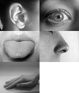
Humans have a multitude of senses. Sight (ophthalmoception), hearing (audioception), taste (gustaoception), smell (olfacoception or olfacception), and touch (tactioception) are the five traditionally recognized senses. The ability to detect other stimuli beyond those governed by these most broadly recognized senses also exists, and these sensory modalities include temperature (thermoception), kinesthetic sense (proprioception), pain (nociception), balance (equilibrioception), vibration (mechanoreception), and various internal stimuli (e.g. the different chemoreceptors for detecting saltand carbon dioxide concentrations in the blood). However, what constitutes a sense is a matter of some debate, leading to difficulties in defining what exactly a distinct sense is, and where the borders between responses to related stimuli lay.
Other animals also have receptors to sense the world around them, with degrees of capability varying greatly between species. Humans have a comparatively weak sense of smell relative to many other mammals while some animals may lack one or more of the traditional five senses. Some animals may also intake and interpret sensory stimuli in very different ways. Some species of animals are able to sense the world in a way that humans cannot, with some species able to sense electrical and magnetic fields, and detect water pressure and currents.
Contents
Definition
A broadly acceptable definition of a sense would be "A system that consists of a group of sensory cell types that responds to a specific physical phenomenon, and that corresponds to a particular group of regions within the brain where the signals are received and interpreted." There is no firm agreement as to the number of senses because of differing definitions of what constitutes a sense.
The senses are frequently divided into exteroceptive and interoceptive:
Non-human animals may possess senses that are absent in humans, such as electroreception and detection of polarized light.
In Buddhist philosophy, Ayatana or "sense-base" includes the mind as a sense organ, in addition to the traditional five. This addition to the commonly acknowledged senses may arise from the psychological orientation involved in Buddhist thought and practice. The mind considered by itself is seen as the principal gateway to a different spectrum of phenomena that differ from the physical sense data. This way of viewing the human sense system indicates the importance of internal sources of sensation and perception that complements our experience of the external world.
Traditional senses
See also: Five wits § The "outward" wits
Sight
Sight or vision (adjectival form: visual/optical) is the capability of the eye(s) to focus and detect images of visible light on photoreceptors in the retina of each eye that generates electrical nerve impulses for varying colors, hues, and brightness. There are two types of photoreceptors: rods and cones. Rods are very sensitive to light, but do not distinguish colors. Cones distinguish colors, but are less sensitive to dim light. There is some disagreement as to whether this constitutes one, two or three senses. Neuroanatomists generally regard it as two senses, given that different receptors are responsible for the perception of color and brightness. Some argue[citation needed] that stereopsis, the perception of depth using both eyes, also constitutes a sense, but it is generally regarded as a cognitive (that is, post-sensory) function of the visual cortex of the brain where patterns and objects in images are recognized and interpreted based on previously learned information. This is called visual memory.
The inability to see is called blindness. Blindness may result from damage to the eyeball, especially to the retina, damage to the optic nerve that connects each eye to the brain, and/or from stroke (infarcts in the brain). Temporary or permanent blindness can be caused by poisons or medications.
People who are blind from degradation or damage to the visual cortex, but still have functional eyes, are actually capable of some level of vision and reaction to visual stimuli but not a conscious perception; this is known as blindsight. People with blindsight are usually not aware that they are reacting to visual sources, and instead just unconsciously adapt their behaviour to the stimulus.
On February 14, 2013 researchers developed a neural implant that gives rats the ability to sense infrared light which for the first time provides living creatures with new abilities, instead of simply replacing or augmenting existing abilities.[3]
Hearing
Hearing or audition (adjectival form: auditory) is the sense of sound perception. Hearing is all about vibration. Mechanoreceptors turn motion into electrical nerve pulses, which are located in the inner ear. Since sound is vibration, propagating through a medium such as air, the detection of these vibrations, that is the sense of the hearing, is a mechanical sense because these vibrations are mechanically conducted from the eardrum through a series of tiny bones to hair-like fibers in the inner ear, which detect mechanical motion of the fibers within a range of about 20 to 20,000 hertz,[4]with substantial variation between individuals. Hearing at high frequencies declines with an increase in age. Inability to hear is called deafness or hearing impairment. Sound can also be detected as vibrations conducted through the body by tactition. Lower frequencies that can be heard are detected this way. Some deaf people are able to determine direction and location of vibrations picked up through the feet.[5]
Taste
Taste (or, the more formal term, gustation; adjectival form: gustatory) is one of the traditional five senses. It refers to the capability to detect the taste of substances such as food, certain minerals, and poisons, etc. The sense of taste is often confused with the "sense" of flavor, which is a combination of taste and smell perception. Flavor depends on odor, texture, and temperature as well as on taste. Humans receive tastes through sensory organs called taste buds, or gustatory calyculi, concentrated on the upper surface of the tongue. There are five basic tastes: sweet, bitter, sour, salty and umami. Other tastes such as calcium[6][7] and free fatty acids[8] may also be basic tastes but have yet to receive widespread acceptance. The inability to taste is called ageusia.
Smell
Smell or olfaction (adjectival form: olfactory) is the other "chemical" sense. Unlike taste, there are hundreds of olfactory receptors (388 according to one source[9]), each binding to a particular molecular feature. Odor molecules possess a variety of features and, thus, excite specific receptors more or less strongly. This combination of excitatory signals from different receptors makes up what we perceive as the molecule's smell. In the brain, olfaction is processed by the olfactory system.Olfactory receptor neurons in the nose differ from most other neurons in that they die and regenerate on a regular basis. The inability to smell is called anosmia. Some neurons in the nose are specialized to detect pheromones.[10]
Touch
Touch or somatosensation (adjectival form: somatic), also called tactition (adjectival form: tactile) ormechanoreception, is a perception resulting from activation of neural receptors, generally in the skin including hair follicles, but also in the tongue, throat, and mucosa. A variety of pressure receptors respond to variations in pressure (firm, brushing, sustained, etc.). The touch sense of itching caused by insect bites or allergies involves special itch-specific neurons in the skin and spinal cord.[11] The loss or impairment of the ability to feel anything touched is called tactileanesthesia. Paresthesia is a sensation of tingling, pricking, or numbness of the skin that may result from nerve damage and may be permanent or temporary.
Non-traditional senses
Balance and acceleration
Main article: Vestibular system
Balance, equilibrioception, or vestibular sense is the sense that allows an organism to sense body movement, direction, and acceleration, and to attain and maintain postural equilibrium and balance. The organ of equilibrioception is the vestibular labyrinthine system found in both of the inner ears. In technical terms, this organ is responsible for two senses of angular momentum acceleration and linear acceleration (which also senses gravity), but they are known together as equilibrioception.
The vestibular nerve conducts information from sensory receptors in three ampulla that sense motion of fluid in threesemicircular canals caused by three-dimensional rotation of the head. The vestibular nerve also conducts information from the utricle and the saccule, which contain hair-like sensory receptors that bend under the weight of otoliths (which are small crystals of calcium carbonate) that provide the inertia needed to detect head rotation, linear acceleration, and the direction of gravitational force.
Temperature
Thermoception is the sense of heat and the absence of heat (cold) by the skin and including internal skin passages, or, rather, the heat flux (the rate of heat flow) in these areas. There are specialized receptors for cold (declining temperature) and for heat. The cold receptors play an important part in the animal's sense of smell, telling wind direction. The heat receptors are sensitive to infrared radiation and can occur in specialized organs, for instance in pit vipers. Thethermoceptors in the skin are quite different from the homeostatic thermoceptors in the brain (hypothalamus), which provide feedback on internal body temperature.
Kinesthetic sense
Proprioception, the kinesthetic sense, provides the parietal cortex of the brain with information on the relative positions of the parts of the body. Neurologists test this sense by telling patients to close their eyes and touch their own nose with the tip of a finger. Assuming proper proprioceptive function, at no time will the person lose awareness of where the hand actually is, even though it is not being detected by any of the other senses. Proprioception and touch are related in subtle ways, and their impairment results in surprising and deep deficits in perception and action.[12]
Pain
Nociception (physiological pain) signals nerve-damage or damage to tissue. The three types of pain receptors are cutaneous (skin), somatic (joints and bones), and visceral (body organs). It was previously believed that pain was simply the overloading of pressure receptors, but research in the first half of the 20th century indicated that pain is a distinct phenomenon that intertwines with all of the other senses, including touch. Pain was once considered an entirely subjective experience, but recent studies show that pain is registered in the anterior cingulate gyrus of the brain.[13] The main function of pain is to attract our attention to dangers and motivate us to avoid them. For example, humans avoid touching a sharp needle, or hot object, or extending an arm beyond a safe limit because it is dangerous, and thus hurts. Without pain, people could do many dangerous things without being aware of the dangers.
Other internal senses
An internal sense also known as interoception[14] is "any sense that is normally stimulated from within the body".[15]These involve numerous sensory receptors in internal organs, such as stretch receptors that are neurologically linked to the brain. Some examples of specific receptors are:
Perception not based on a specific sensory organ
Time
Chronoception refers to how the passage of time is perceived and experienced. Although the sense of time is not associated with a specific sensory system, the work of psychologists and neuroscientists indicates that human brains do have a system governing the perception of time,[17][18] composed of a highly distributed system involving the cerebral cortex,cerebellum and basal ganglia. One particular component, the suprachiasmatic nucleus, is responsible for the circadian (or daily) rhythm, while other cell clusters appear to be capable of shorter-range (ultradian) timekeeping.
Non-human senses
Analogous to human senses
Other living organisms have receptors to sense the world around them, including many of the senses listed above for humans. However, the mechanisms and capabilities vary widely.
Smell
Most non-human mammals have a much keener sense of smell than humans, although the mechanism is similar. Sharkscombine their keen sense of smell with timing to determine the direction of a smell. They follow the nostril that first detected the smell.[19] Insects have olfactory receptors on their antennae.
Vomeronasal organ
Many animals (salamanders, reptiles, mammals) have a vomeronasal organ[20] that is connected with the mouth cavity. In mammals it is mainly used to detect pheromones of marked territory, trails, and sexual state. Reptiles like snakes andmonitor lizards make extensive use of it as a smelling organ by transferring scent molecules to the vomeronasal organ with the tips of the forked tongue. In reptiles the vomeralnasal organ is commonly referred to as Jacobsons organ. In mammals, it is often associated with a special behavior called flehmen characterized by uplifting of the lips. The organ is vestigial in humans, because associated neurons have not been found that give any sensory input in humans.[21]
Taste
Flies and butterflies have taste organs on their feet, allowing them to taste anything they land on. Catfish have taste organs across their entire bodies, and can taste anything they touch, including chemicals in the water.[22]
Vision
Cats have the ability to see in low light, which is due to muscles surrounding their irises–which contract and expand their pupils–as well as to the tapetum lucidum, a reflective membrane that optimizes the image. Pit vipers, pythons and some boashave organs that allow them to detect infrared light, such that these snakes are able to sense the body heat of their prey. The common vampire bat may also have an infrared sensor on its nose.[23] It has been found that birds and some other animals are tetrachromats and have the ability to see in the ultraviolet down to 300 nanometers. Bees and dragonflies[24]are also able to see in the ultraviolet. Mantis shrimps can perceive both polarized light and multispectral images and are dodecachromats.[25]
Balance
Many invertebrates have a statocyst, which is a sensor for acceleration and orientation that works very differently from the mammalian's semi-circular canals.
Sensing gravity
Some plants (such as mustard) have genes that are necessary for the plant to sense the direction of gravity. If these genes are disabled by a mutation, a plant cannot grow upright.[26]
Not analogous to human senses
In addition, some animals have senses that humans do not, including the following:
Echolocation
Main article: Animal echolocation
Certain animals, including bats and cetaceans, have the ability to determine orientation to other objects through interpretation of reflected sound (like sonar). They most often use this to navigate through poor lighting conditions or to identify and track prey. There is currently an uncertainty whether this is simply an extremely developed post-sensory interpretation of auditory perceptions or it actually constitutes a separate sense. Resolution of the issue will require brain scans of animals while they actually perform echolocation, a task that has proven difficult in practice.
Blind people report they are able to navigate and in some cases identify an object by interpreting reflected sounds (especially their own footsteps), a phenomenon known as human echolocation.
Electroreception
Electroreception (or electroception) is the ability to detect electric fields. Several species of fish, sharks, and rays have the capacity to sense changes in electric fields in their immediate vicinity. For cartilaginous fish this occurs through a specialized organ called the Ampullae of Lorenzini. Some fish passively sense changing nearby electric fields; some generate their own weak electric fields, and sense the pattern of field potentials over their body surface; and some use these electric field generating and sensing capacities for social communication. The mechanisms by which electroceptive fish construct a spatial representation from very small differences in field potentials involve comparisons of spike latencies from different parts of the fish's body.
The only orders of mammals that are known to demonstrate electroception are the dolphin and monotreme orders. Among these mammals, the platypus[27] has the most acute sense of electroception.
A dolphin can detect electric fields in water using electroreceptors in vibrissal crypts arrayed in pairs on its snout and which evolved from whisker motion sensors.[28] These electroreceptors can detect electric fields as weak as 4.6 microvolts per centimeter, such as those generated by contracting muscles and pumping gills of potential prey. This permits the dolphin to locate prey from the seafloor where sediment limits visibility and echolocation.
Body modification enthusiasts have experimented with magnetic implants to attempt to replicate this sense.[29] However, in general humans (and it is presumed other mammals) can detect electric fields only indirectly by detecting the effect they have on hairs. An electrically charged balloon, for instance, will exert a force on human arm hairs, which can be felt through tactition and identified as coming from a static charge (and not from wind or the like). This is not electroreception, as it is a post-sensory cognitive action.
Magnetoception
Magnetoception (or magnetoreception) is the ability to detect the direction one is facing based on the Earth's magnetic field. Directional awareness is most commonly observed in birds.[30][31][32] It has also been observed in insects such as bees. Although there is no dispute that this sense exists in many avians (it is essential to the navigational abilities of migratory birds), it is not a well-understood phenomenon.[33] One study has found that cattle make use of magnetoception, as they tend to align themselves in a north-south direction.[34] Magnetotactic bacteria build miniature magnets inside themselves and use them to determine their orientation relative to the Earth's magnetic field.[35][36] The question of how useful magnetoception may be to human beings is subject of ongoing research.[37]
Other
Plant senses
By using a variety of sense receptors, plants sense light, gravity, temperature, humidity, chemical substances, chemical gradients, reorientation, magnetic fields, infections, damage to their tissues and mechanical pressure. The absence of a nervous system notwithstanding, plants interpret and respond to these stimuli by a variety of hormonal and cell-to-cell communication pathways that result in movement, morphological changes and physiological state alterations at the organism level, that is, result in plant behavior. Such physiological and cognitive functions are generally not believed to give rise to mental phenomena or qualia, however, as these are typically considered the product of nervous system activity. The emergence of mental phenomena from the activity of systems functionally or computationally analogous to that of nervous systems is, however, a hypothetical possibility explored by some schools of thought in the philosophy of mind field, such as functionalism and computationalism.
Culture
In the time of William Shakespeare, there were commonly reckoned to be five witsor five senses.[38] At that time, the words "sense" and "wit" were synonyms,[38] so the senses were known as the five outward wits.[39][40] This traditional concept of five senses is common today.
The traditional five senses are enumerated as the "five material faculties" (pañcannaṃ indriyānaṃ avakanti) in Buddhist literature. They appear in allegorical representation as early as in the Katha Upanishad (roughly 6th century BC), as five horses drawing the "chariot" of the body, guided by the mind as "chariot driver".
Depictions of the five traditional senses as allegory became a popular subject for seventeenth-century artists, especially among Dutch and Flemish Baroque painters. A typical example is Gérard de Lairesse's Allegory of the Five Senses (1668), in which each of the figures in the main group alludes to a sense: Sight is the reclining boy with a convex mirror, hearing is the cupid-like boy with a triangle, smell is represented by the girl with flowers, taste is represented by the woman with the fruit, and touch is represented by the woman holding the bird.
In Tamil literature, the Tolkāppiyam is said to be the first written text in the world to describe six senses which related to external body parts. One of its verses says "beings with one sense are those that have the sense of TOUCH. Beings with two senses are those that have the sense of TASTE along with the above. Beings with three senses, have sense of SMELL in addition. Beings with four senses, have sense of SIGHT, along with the above. Beings with five senses, have sense of HEARING, in addition. The beings with six senses, have a MIND, along with the above."[41][42]
Human brain
This article is about features specific to the human brain. For basic information about brains, see Brain.
| Human brain | |
|---|---|

Human brain and skull
| |

| |
| Details | |
| Latin | Cerebrum |
| Greek | ἐγκέφαλος (enképhalos) μυαλό (myaló) |
| Precursor | Neural tube |
| System | Central nervous system |
| Internal carotid arteries, vertebral arteries | |
| Internal jugular vein, cerebral veins, external veins, basal vein,terminal vein, choroid vein,cerebellar veins | |
| Anatomical terminology | |
The human brain is the main organ of the human nervous system. It is located in the head, protected by the skull. It has the same general structure as the brains of other mammals, but with a more developed cerebral cortex. Large animals such as whales and elephants have larger brains in absolute terms, but when measured using a measure of relative brain size, which compensates for body size, the quotient for the human brain is almost twice as large as that of a bottlenose dolphin, and three times as large as that of a chimpanzee. Much of the size of the human brain comes from the cerebral cortex, especially the frontal lobes, which are associated with executive functions such as self-control, planning, reasoning, and abstract thought. The area of the cerebral cortex devoted to vision, the visual cortex, is also greatly enlarged in humans compared to other animals.
The human cerebral cortex is a thick layer of neural tissue that covers most of the brain. This layer is folded in a way that increases the amount of surface that can fit into the volume available. The pattern of folds is similar across individuals, although there are many small variations. The cortex is divided into four lobes – the frontal lobe, parietal lobe, temporal lobe, and occipital lobe. (Some classification systems also include a limbic lobe and treat the insular cortex as a lobe.) Within each lobe are numerous cortical areas, each associated with a particular function, including vision, motor control, andlanguage. The left and right sides of the cortex are broadly similar in shape, and most cortical areas are replicated on both sides. Some areas, though, show strong lateralization, particularly areas that are involved in language. In most people, the left hemisphere is dominant for language, with the right hemisphere playing only a minor role. There are other functions, such asvisual-spatial ability, for which the right hemisphere is usually dominant.
Despite being protected by the thick bones of the skull, suspended incerebrospinal fluid, and isolated from the bloodstream by the blood–brain barrier, the human brain is susceptible to damage and disease. The most common forms of physical damage are closed head injuries such as a blow to the head, a stroke, or poisoning by a variety of chemicals which can act asneurotoxins, such as ethanol alcohol. Infection of the brain, though serious, is rare because of the biological barriers which protect it. The human brain is also susceptible to degenerative disorders, such as Parkinson's disease, andAlzheimer's disease, (mostly as the result of aging) and multiple sclerosis. A number of psychiatric conditions, such as schizophrenia and clinical depression, are thought to be associated with brain dysfunctions, although the nature of these is not well understood. The brain can also be the site of brain tumors and these can be benign or malignant.
Scientifically, the techniques that are used to study the human brain differ in important ways from those that are used to study the brains of other mammals. On the one hand, invasive techniques such as inserting electrodes into the brain, or disabling parts of the brain in order to examine the effect on behavior, are used with non-human species, but for ethical reasons, are generally not performed with humans. On the other hand, humans are the only subjects who can respond to complex verbal instructions. Thus, it is often possible to use non-invasive techniques such as functional neuroimaging or EEG recording more productively with humans than with non-humans. Furthermore, some of the most important topics, such as language, can hardly be studied at all except in humans. In many cases, human and non-human studies form essential complements to each other. Individual brain cells (except where tissue samples are taken for biopsy for suspected brain tumors) can only be studied in non-humans; complex cognitive tasks can only be studied in humans. Combining the two sources of information to yield a complete functional understanding of the human brain is an ongoing challenge for neuroscience.
Contents
Structure
The adult human brain weighs on average about 1.3–1.4 kg (2.9–3.1 lb), or about 2% of total body weight,[1][2] with a volume of around 1130 cubic centimetres (cm3) in women and 1260 cm3 in men, although there is substantial individual variation.[3] Neurological differences between the sexes have not been shown to correlate in any simple way with IQ or other measures of cognitive performance.[4]
The human brain is composed of neurons, glial cells, and blood vessels. The number of neurons, according to array tomography, has been shown to be on average about 86 billion in the adult male human brain with a roughly equal number of non-neuronal cells. Out of these, 16 billion (or 19% of all brain neurons) are located in the cerebral cortex (including subcortical white matter), 69 billion (or 80% of all brain neurons) are in the cerebellum, and fewer than 1% of all brain neurons are located in the rest of the brain.[5]
The cerebral hemispheres (the cerebrum) form the largest part of the human brain and are situated above other brain structures. They are covered with a cortical layer (the cerebral cortex) which has a convoluted topography.[6] Underneath the cerebrum lies the brainstem, resembling a stalk on which the cerebrum is attached. At the rear of the brain, beneath the cerebrum and behind the brainstem, is the cerebellum, a structure with a horizontally furrowed surface, the cerebellar cortex, that makes it look different from any other brain area. The same structures are present in other mammals, although they vary considerably in relative size. As a rule, the smaller the cerebrum, the less convoluted the cortex. The cortex of a rat or mouse is almost perfectly smooth. The cortex of a dolphin or whale, on the other hand, is more convoluted than the cortex of a human.
The living brain is very soft, having a consistency similar to soft gelatin or soft tofu. Although referred to as grey matter, the live cortex is pinkish-beige in color and slightly off-white in the interior.
General features
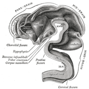
The human brain has many properties that are common to allvertebrate brains, including a basic division into three parts called theforebrain, midbrain, and hindbrain, with interconnected fluid-filledventricles, and a set of generic vertebrate brain structures including themedulla oblongata and pons of the brainstem, the cerebellum, optic tectum, thalamus, hypothalamus, basal ganglia, olfactory bulb, and many others.
As a mammalian brain, the human brain has special features that are common to all mammalian brains, most notably a six-layered cerebral cortex and a set of structures associated with it, including thehippocampus and amygdala. All vertebrates have a forebrain whose upper surface is covered with a layer of neural tissue called the pallium, but in all except mammals the pallium has a relatively simple three-layered cell structure. In mammals it has a much more complex six-layered cell structure, and is given a different name, the cerebral cortex. The hippocampus and amygdala also originate from the pallium, but are much more complex in mammals than in other vertebrates.
As a primate brain, the human brain has a much larger cerebral cortex, in proportion to body size, than most mammals, and a very highly developed visual system. The shape of the brain within the skull is also altered somewhat as a consequence of the upright position in which primates hold their heads.
As a hominid brain, the human brain is substantially enlarged even in comparison to the brain of a typical monkey. The sequence of evolution from Australopithecus (four million years ago) to Homo sapiens (modern man) was marked by a steady increase in brain size, particularly in the frontal lobes, which are associated with a variety of high-level cognitive functions.
Humans and other primates have some differences in gene sequence, and genes are differentially expressed in many brain regions. The functional differences between the human brain and the brains of other animals also arise from many gene–environment interactions.[7]
Cerebral cortex
The dominant feature of the human brain is corticalization. The cerebral cortex in humans is so large that it overshadows every other part of the brain.[9] A few subcortical structures show alterations reflecting this trend. The cerebellum, for example, has a medial zone connected mainly to subcortical motor areas, and a lateral zone connected primarily to the cortex. In humans the lateral zone takes up a much larger fraction of the cerebellum than in most other mammalian species. Corticalization is reflected in function as well as structure. In a rat, surgical removal of the entire cerebral cortex leaves an animal that is still capable of walking around and interacting with the environment.[10] In a human, comparable cerebral cortex damage produces a permanent state of coma. The amount of association cortex, relative to the other two categories of sensory and motor, increases dramatically as one goes from simpler mammals, such as the rat and the cat, to more complex ones, such as the chimpanzee and the human.[11] A gene present in the human genome but not in the chimpanzee : ArhGAP11B seems to play a major role in "corticalization. ArhGAP11B and human encephalisation The cerebral cortex is essentially a sheet of neural tissue, folded in a way that allows a large surface area to fit within the confines of the skull. When unfolded, each cerebral hemisphere has a total surface area of about 1.3 square feet (0.12 m2).[12] Each cortical ridge is called a gyrus, and each groove or fissure separating one gyrus from another is called a sulcus.
Cortical divisions
The cerebral cortex is nearly symmetrical with left and right hemispheres that are approximate mirror images of each other. Each hemisphere is conventionally divided into four "lobes", the frontal lobe,parietal lobe, occipital lobe, and temporal lobe. With one exception, this division into lobes does not derive from the structure of the cortex, though the lobes are named after the bones of the skull that overlie them, the frontal bone, parietal bone, temporal bone, and occipital bone. The borders between lobes lie beneath the sutures that link the skull bones together. The exception is the border between the frontal and parietal lobes, which lies behind the corresponding suture; instead it follows the anatomical boundary of the central sulcus, a deep fold in the brain's structure where the primary somatosensory cortex and primary motor cortex meet.
Because of the arbitrary way most of the borders between lobes are demarcated, they have little functional significance. With the exception of the occipital lobe, a small area that is entirely dedicated to vision, each of the lobes contains a variety of brain areas that have minimal functional relationship. The parietal lobe, for example, contains areas involved in somatosensation, hearing, language, attention, and spatial cognition. In spite of this heterogeneity, the division into lobes is convenient for reference. The main functions of the frontal lobe are to control attention, abstract thinking, behavior, problem solving tasks, and physical reactions and personality.[13] The occipital lobe is the smallest lobe; its main functions are visual reception, visual-spatial processing, movement, and color recognition.[14] The temporal lobe controls auditory and visual memories, language, and some hearing and speech.[13]
Although there are enough variations in the shape and placement of gyri and sulci(cortical folds) to make every brain unique, most human brains show sufficiently consistent patterns of folding that allow them to be named. Many of the gyri and sulci are named according to the location on the lobes or other major folds on the cortex. These include:
Functional divisions
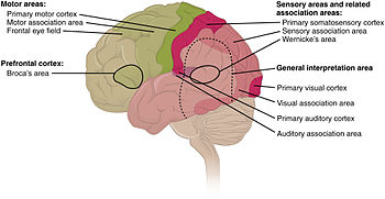
Researchers who study the functions of the cortex divide it into three functional categories of regions. One consists of theprimary sensory areas, which receive signals from the sensory nerves and tracts by way of relay nuclei in the thalamus. Primary sensory areas include the visual area of the occipital lobe, the auditory area in parts of the temporal lobe andinsular cortex, and the somatosensory cortex in the parietal lobe. A second category is the primary motor cortex, which sends axons down to motor neurons in the brainstem and spinal cord.[15] This area occupies the rear portion of the frontal lobe, directly in front of the somatosensory area. The third category consists of the remaining parts of the cortex, which are called the association areas. These areas receive input from the sensory areas and lower parts of the brain and are involved in the complex processes of perception, thought, and decision-making.[16]
Cytoarchitecture
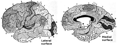
Different parts of the cerebral cortex are involved in different cognitive and behavioral functions. The differences show up in a number of ways: the effects of localized brain damage, regional activity patterns exposed when the brain is examined using functional imaging techniques, connectivity with subcortical areas, and regional differences in the cellular architecture of the cortex. Neuroscientists describe most of the cortex—the part they call the neocortex—as having six layers, but not all layers are apparent in all areas, and even when a layer is present, its thickness and cellular organization may vary. Scientists have constructed maps of cortical areason the basis of variations in the appearance of the layers as seen with a microscope. One of the most widely used schemes came from Korbinian Brodmann, who split the cortex into 51 different areas and assigned each a number (many of theseBrodmann areas have since been subdivided). For example, Brodmann area 1 is the primary somatosensory cortex, Brodmann area 17 is the primary visual cortex, and Brodmann area 25 is the anterior cingulate cortex.[17]
Topography
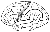
Many of the brain areas Brodmann defined have their own complex internal structures. In a number of cases, brain areas are organized into "topographic maps", where adjoining bits of the cortex correspond to adjoining parts of the body, or of some more abstract entity. A simple example of this type of correspondence is the primary motor cortex, a strip of tissue running along the anterior edge of the central sulcus, shown in the image to the right. Motor areas innervating each part of the body arise from a distinct zone, with neighboring body parts represented by neighboring zones. Electrical stimulation of the cortex at any point causes a muscle-contraction in the represented body part. This "somatotopic" representation is not evenly distributed, however. The head, for example, is represented by a region about three times as large as the zone for the entire back and trunk. The size of any zone correlates to the precision of motor control and sensory discrimination possible. The areas for the lips, fingers, and tongue are particularly large, considering the proportional size of their represented body parts.
In visual areas, the maps are retinotopic—that is, they reflect the topography of the retina, the layer of light-activated neurons lining the back of the eye. In this case too the representation is uneven: the fovea—the area at the center of the visual field—is greatly overrepresented compared to the periphery. The visual circuitry in the human cerebral cortex contains several dozen distinct retinotopic maps, each devoted to analyzing the visual input stream in a particular way. The primary visual cortex (Brodmann area 17), which is the main recipient of direct input from the visual part of the thalamus, contains many neurons that are most easily activated by edges with a particular orientation moving across a particular point in the visual field. Visual areas farther downstream extract features such as color, motion, and shape.
In auditory areas, the primary map is tonotopic. Sounds are parsed according to frequency (i.e., high pitch vs. low pitch) by subcortical auditory areas, and this parsing is reflected by the primary auditory zone of the cortex. As with the visual system, there are a number of tonotopic cortical maps, each devoted to analyzing sound in a particular way.
Within a topographic map there can sometimes be finer levels of spatial structure. In the primary visual cortex, for example, where the main organization is retinotopic and the main responses are to moving edges, cells that respond to different edge-orientations are spatially segregated from one another.
Development
Main article: Neural development in humans
Further information: Human brain development timeline
During the first three weeks of gestation, the human embryo's ectoderm forms a thickened strip called the neural plate. Theneural plate then folds and closes to form the neural tube. This tube flexes as it grows, forming the crescent-shaped cerebral hemispheres at the head, and the cerebellum and pons towards the tail.
Function
Cognition
Understanding the mind–body problem – the relationship between the brain and the mind – is a significant challenge both philosophically and scientifically. It is very difficult to imagine how mental activities such as thoughts and emotions could be implemented by physical structures such as neurons and synapses, or by any other type of physical mechanism. This difficulty was expressed by Gottfried Leibniz in an analogy known as Leibniz's Mill:
Doubt about the possibility of a mechanistic explanation of thought drove René Descartes, and most of humankind along with him, to dualism: the belief that the mind exists independently of the brain.[19] There has always, however, been a strong argument in the opposite direction. There is clear empirical evidence that physical manipulations of, or injuries to, the brain (for example by drugs or by lesions, respectively) can affect the mind in potent and intimate ways.[20] For example, a person suffering from Alzheimer's disease – a condition that causes physical damage to the brain – also experiences a compromised mind. Similarly, someone who has taken a psychedelic drug may temporarily lose their sense of personal identity (ego death) or experience profound changes to their perception and thought processes. Likewise, a patient withepilepsy who undergoes cortical stimulation mapping with electrical brain stimulation would also, upon stimulation of his or her brain, experience various complex feelings, hallucinations, memory flashbacks, and other complex cognitive, emotional, or behavioral phenomena.[21] Following this line of thinking, a large body of empirical evidence for a close relationship between brain activity and mental activity has led most neuroscientists and contemporary philosophers to be materialists, believing that mental phenomena are ultimately the result of, or reducible to, physical phenomena.[22]
Lateralization
Main article: Lateralization of brain function
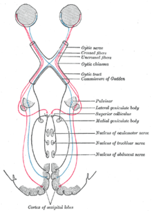
Each hemisphere of the brain interacts primarily with one half of the body, but for reasons that are unclear, the connections are crossed: the left side of the brain interacts with the right side of the body, and vice versa.[citation needed] Motor connections from the brain to the spinal cord, and sensory connections from the spinal cord to the brain, both cross the midline at the level of the brainstem. Visual input follows a more complex rule: the optic nerves from the two eyes come together at a point called the optic chiasm, and half of the fibers from each nerve split off to join the other. The result is that connections from the left half of the retina, in both eyes, go to the left side of the brain, whereas connections from the right half of the retina go to the right side of the brain. Because each half of the retina receives light coming from the opposite half of the visual field, the functional consequence is that visual input from the left side of the world goes to the right side of the brain, and vice versa. Thus, the right side of the brain receives somatosensory input from the left side of the body, and visual input from the left side of the visual field—an arrangement that presumably is helpful for visuomotor coordination.
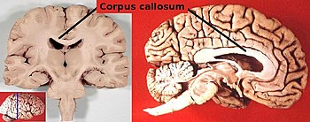
The two cerebral hemispheres are connected by a very large nerve bundle (the largest white matter structure in the brain) called the corpus callosum, which crosses the midline above the level of the thalamus.[23] There are also two much smaller connections, the anterior commissure and hippocampal commissure, as well as many subcortical connections that cross the midline. The corpus callosum is the main avenue of communication between the two hemispheres, though. It connects each point on the cortex to the mirror-image point in the opposite hemisphere, and also connects to functionally related points in different cortical areas.
In most respects, the left and right sides of the brain are symmetrical in terms of function. For example, the counterpart of the left-hemisphere motor area controlling the right hand is the right-hemisphere area controlling the left hand. There are, however, several very important exceptions, involving language and spatial cognition. In most people, the left hemisphere is "dominant" for language: a stroke that damages a key language area in the left hemisphere can leave the victim unable to speak or understand, whereas equivalent damage to the right hemisphere would cause only minor impairment to language skills.
A substantial part of current understanding of the interactions between the two hemispheres has come from the study of "split-brain patients"—people who underwent surgical transection of the corpus callosum in an attempt to reduce the severity of epileptic seizures. These patients do not show unusual behavior that is immediately obvious, but in some cases can behave almost like two different people in the same body, with the right hand taking an action and then the left hand undoing it. Most of these patients, when briefly shown a picture on the right side of the point of visual fixation, are able to describe it verbally, but when the picture is shown on the left, are unable to describe it, but may be able to give an indication with the left hand of the nature of the object shown.
Language
Main article: Language processing in the brain
The study of how language is represented, processed, andacquired by the brain is neurolinguistics, which is a large multidisciplinary field drawing from cognitive neuroscience,cognitive linguistics, and psycholinguistics. This field originated from the 19th-century discovery that damage to different parts of the brain appeared to cause different symptoms: physicians noticed that individuals with damage to a portion of the leftinferior frontal gyrus now known as Broca's area had difficulty in producing language (aphasia of speech), whereas those with damage to a region in the left superior temporal gyrus, now known as Wernicke's area, had difficulty in understanding it.[24]
Since then, there has been substantial debate over what linguistic processes these and other parts of the brain subserve,[25] and although Broca's and Wernicke's areas have traditionally been associated with language functions, they may also be involved in certain non-speech functions.[citation needed] There is also debate over whether or not there even is a strong one-to-one relationship between brain regions and language functions that emerges during neocortical development.[26] More recently, research on language has increasingly used more modern methods including electrophysiology and functional neuroimaging, to examine how language processing occurs. In the study of natural language, a dedicated network of language development has been identified as crucially involving Broca's area.[27][28]
Metabolism
The brain consumes up to twenty percent of the energy used by the human body, more than any other organ.[29] Brain metabolism normally relies upon blood glucose as an energy source, but during times of low glucose (such as fasting, exercise, or limited carbohydrate intake), the brain will use ketone bodies for fuel with a smaller need for glucose. The brain can also utilize lactate during exercise.[30] Long-chain fatty acids cannot cross the blood–brain barrier, but the liver can break these down to produce ketones. However the medium-chain fatty acids octanoicand heptanoic acids can cross the barrier and be used by the brain.[31][32][33] The brain stores glucose in the form of glycogen, albeit in significantly smaller amounts than that found in the liver or skeletal muscle.[34]
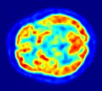
Although the human brain represents only 2% of the body weight, it receives 15% of the cardiac output, 20% of total body oxygen consumption, and 25% of total body glucose utilization.[35]The need to limit body weight has led to selection for a reduction of brain size in some species, such as bats, who need to be able to fly.[36] The brain mostly uses glucose for energy, and deprivation of glucose, as can happen in hypoglycemia, can result in loss of consciousness. The energy consumption of the brain does not vary greatly over time, but active regions of the cortex consume somewhat more energy than inactive regions: this fact forms the basis for the functional brain imaging methods PET and fMRI.[37] These are nuclear medicine imaging techniques which produce a three-dimensional image of metabolic activity.
Clinical significance
Clinically, death is defined as an absence of brain activity as measured by EEG. Injuries to the brain tend to affect large areas of the organ, sometimes causing major deficits in intelligence, memory, personality, and movement. Head trauma caused, for example, by vehicular or industrial accidents, is a leading cause of death in youth and middle age. In many cases, more damage is caused by resultant edema than by the impact itself. Stroke, caused by the blockage or rupturing of blood vessels in the brain, is another major cause of death from brain damage.
Other problems in the brain can be more accurately classified as diseases. Neurodegenerative diseases, such asAlzheimer's disease, Parkinson's disease, Huntington's disease and motor neuron diseases are caused by the gradual death of individual neurons, leading to diminution in movement control, memory, and cognition. There are five motor neuron diseases, the most common of which is amyotrophic lateral sclerosis (ALS).
Some infectious diseases affecting the brain are caused by viruses and bacteria. Infection of the meninges, the membranes that cover the brain, can lead to meningitis. Bovine spongiform encephalopathy (also known as "mad cow disease") is deadly in cattle and humans and is linked to prions. Kuru is a similar prion-borne degenerative brain disease affecting humans, (endemic only to Papua New Guinea tribes). Both are linked to the ingestion of neural tissue, and may explain the tendency in human and some non-human species to avoid cannibalism. Viral or bacterial causes have been reported inmultiple sclerosis, and are established causes of encephalopathy, and encephalomyelitis.
Brain tumors both benign and malignant can form. These can either originate in the cerebral tissue or in the meninges. The most common are those growths that affect the glial cells known as gliomas. (This term has been extended to include all primary brain tumors.)[38] Secondary cancers can form in the brain as a result of brain metastasis.
Mental disorders, such as clinical depression, schizophrenia, bipolar disorder and post-traumatic stress disorder may involve particular patterns of neuropsychological functioning related to various aspects of mental and somatic function. These disorders may be treated by psychotherapy, psychiatric medication, social intervention and personal recovery work orcognitive behavioural therapy; the underlying issues and associated prognoses vary significantly between individuals.
Many brain disorders are congenital, occurring during development. Tay-Sachs disease, fragile X syndrome, and Down syndrome are all linked to genetic and chromosomal errors. Many other syndromes, such as the intrinsic circadian rhythmdisorders, are suspected to be congenital as well. Normal development of the brain can be altered by genetic factors, drug use, nutritional deficiencies, and infectious diseases during pregnancy.
Epileptic, and non-epileptic seizures can cause cognitive impairment when the seizures become widespread, occur repeatedly in the same brain area or last for too long. Seizures can be assessed using EEG and various medical imagingtechniques. They can sometimes be treated using anticonvulsant drugs and certain neurosurgical procedures and auxiliary treatments may also be used.
Effects of brain damage
A key source of information about the function of brain regions is the effects of damage to them.[39] In humans, strokes have long provided a "natural laboratory" for studying the effects of brain damage. Most strokes result from a blood clot lodging in the brain and blocking the local blood supply, causing damage or destruction of nearby brain tissue: the range of possible blockages is very wide, leading to a great diversity of stroke symptoms. Analysis of strokes is limited by the fact that damage often crosses into multiple regions of the brain, not along clear-cut borders, making it difficult to draw firm conclusions.
Transient ischemic attacks (TIAs) are mini-strokes that can cause sudden dimming or loss of vision (including amaurosis fugax), speech impairment ranging from slurring to dysarthria or aphasia, and mental confusion. But unlike a stroke, the symptoms of a TIA can resolve within a few minutes or 24 hours. Brain injury may still occur in a TIA lasting only a few minutes.[40][41] A silent stroke or silent cerebral infarct (SCI) differs from a TIA in that there are no immediately observable symptoms. An SCI may still cause long lasting neurological dysfunction affecting such areas as mood, personality, and cognition. An SCI often occurs before or after a TIA or major stroke.[42]
Electrodes and magnetic fields
By placing electrodes on the scalp, it is possible to record the summed electrical activity of the cortex using a methodology known as electroencephalography (EEG).[43] EEG records average neuronal activity from the cerebral cortex and can detect changes in activity over large areas but with low sensitivity for sub-cortical activity. EEG recordings are sensitive enough to detect tiny electrical impulses lasting only a few milliseconds. Most EEG devices have good temporal resolution, but low spatial resolution.
Electrodes can also be placed directly on the surface of the brain (usually during surgical procedures that require removal of part of the skull). This technique, called electrocorticography (ECoG), offers finer spatial resolution than electroencephalography, but is very invasive. In addition to measuring the electric field directly via electrodes placed over the skull, it is possible to measure the magnetic field that the brain generates using a method known asmagnetoencephalography (MEG).[44] This technique also has good temporal resolution like EEG but with much better spatial resolution. The greatest disadvantage of MEG is that, because the magnetic fields generated by neural activity are very subtle, the neural activity must be relatively close to the surface of the brain to detect its magnetic field. MEGs can only detect the magnetic signatures of neurons located in the depths of cortical folds (sulci) that have dendrites oriented in a way that produces a field.
Imaging
Further information: Brain mapping and Outline of brain mapping
Neuroscientists, along with researchers from allied disciplines, study how the human brain works. Such research has expanded considerably in recent decades. The "Decade of the Brain", an initiative of the United States Government in the 1990s, is considered to have marked much of this increase in research.[45] It has been followed in 2013 by the BRAIN Initiative.
Information about the structure and function of the human brain comes from a variety of experimental methods. Most information about the cellular components of the brain and how they work comes from studies of animal subjects, using techniques described in the brain article. Some techniques, however, are used mainly in humans, and therefore are described here.
Structural and functional imaging
Main article: Neuroimaging
There are several methods for detecting brain activity changes using three-dimensional imaging of local changes in blood flow. The older methods are SPECTand PET, which depend on injection of radioactive tracers into the bloodstream. A newer method, functional magnetic resonance imaging (fMRI), has considerably better spatial resolution and involves no radioactivity.[46]Using the most powerful magnets currently available, fMRI can localize brain activity changes to regions as small as one cubic millimeter. The downside is that the temporal resolution is poor: when brain activity increases, the blood flow response is delayed by 1–5 seconds and lasts for at least 10 seconds. Thus, fMRI is a very useful tool for learning which brain regions are involved in a given behavior, but gives little information about the temporal dynamics of their responses. A major advantage for fMRI is that, because it is non-invasive, it can readily be used on human subjects.
Another new non-invasive functional imaging method is functional near-infrared spectroscopy.
Evolution
In the course of evolution of the Homininae, the human brain has grown in volume from about 600 cm3 in Homo habilis to about 1500 cm3 in Homo sapiens neanderthalensis. Subsequently, there has been a shrinking over the past 28,000 years. The male brain has decreased from 1,500 cm3 to 1,350 cm3 while the female brain has shrunk by the same relative proportion.[47] For comparison, Homo erectus, a relative of humans, had a brain size of 1,100 cm3. However, the little Homo floresiensis, with a brain size of 380 cm3, a third of that of their proposed ancestor H. erectus, used fire, hunted, and made stone tools at least as sophisticated as those of H. erectus.[48] In spite of significant changes in social capacity, there has been very little change in brain size fromNeanderthals to the present day.[49] "As large as you need and as small as you can" has been said to summarize the opposite evolutionary constraints on human brain size.[50][51]Changes in the size of the human brain during evolution have been reflected in changes in the ASPM and microcephalin genes.[52]
Studies tend to indicate small to moderate correlations (averaging around 0.3 to 0.4) between brain volume and IQ.[53] The most consistent associations are observed within the frontal, temporal, and parietal lobes, the hippocampi, and the cerebellum, but these only account for a relatively small amount of variance in IQ, which itself has only a partial relationship to general intelligence and real-world performance.[54][55][full citation needed] One study indicated that in humans, fertility and intelligence tend to be negatively correlated—that is to say, the more intelligent, as measured by IQ, exhibit a lower total fertility rate than the less intelligent. According to the model, the present rate of decline is predicted to be 1.34 IQ points per decade.[56]
Nerve
This article is about neural pathways of the peripheral nervous system. For other uses, see Nerve (disambiguation).
| Nerve | |
|---|---|

Nerves (yellow) in the arm
| |
| Details | |
| Latin | nervus |
| Anatomical terminology | |
A nerve is an enclosed, cable-like bundle of axons (the long, slender projections of neurons) in the peripheral nervous system. A nerve provides a common pathway for the electrochemical nerve impulses that are transmitted along each of the axons to peripheral organs.
In the central nervous system, the analogous structures are known astracts.[1][2] Neurons are sometimes called nerve cells, though this term is potentially misleading since many neurons do not form nerves, and nerves also include non-neuronal Schwann cells that coat the axons in myelin.
Each nerve is a cordlike structure that contains many axons, also called nerve fibres. Within a nerve, each axon is surrounded by a layer of connective tissue called the endoneurium. The axons are bundled together into groups calledfascicles, and each fascicle is wrapped in a layer of connective tissue called the perineurium. Finally, the entire nerve is wrapped in a layer of connective tissue called the epineurium.
Contents
Anatomy
Nerves are categorized into three groups based on the direction that signals are conducted:
Nerves can be categorized into two groups based on where they connect to the central nervous system:
Each nerve is covered externally by a dense sheath of connective tissue, theepineurium. Underlying this is a layer of flat cells, the perineurium, which forms a complete sleeve around a bundle of axons. Perineurial septae extend into the nerve and subdivide it into several bundles of fibres. Surrounding each such fibre is theendoneurium. This forms an unbroken tube from the surface of the spinal cord to the level where the axon synapses with its muscle fibres, or ends in sensory receptors. The endoneurium consists of an inner sleeve of material called theglycocalyx and an outer, delicate, meshwork of collagen fibres.[2] Nerves are bundled along with blood vessels, since the neurons of a nerve have fairly high energy requirements.
Within the endoneurium, the individual nerve fibres are surrounded by a low protein liquid called endoneurial fluid. This acts in a similar way to the cerebrospinal fluid in the central nervous system and constitutes a blood-nerve barrier similar to theblood-brain barrier.[3] Molecules are thereby prevented from crossing the blood into the endoneurial fluid. During the development of nerve edema from nerve irritation or (injury), the amount of endoneurial fluid may increase at the site of irritation. This increase in fluid can be visualized usingmagnetic resonance neurography, and thus MR neurography can identify nerve irritation and/or injury.
Physiology
A nerve conveys information in the form of electrochemical impulses (known as nerve impulses or action potentials) carried by the individual neurons that make up the nerve. These impulses are extremely fast, with some myelinated neurons conducting at speeds up to 120 m/s. The impulses travel from one neuron to another by crossing a synapse, the message is converted from electrical to chemical and then back to electrical.[1][2]
Nerves can be categorized into two groups based on function:
Clinical significance
Cancer can spread by invading the spaces around nerves. This is particularly common in head and neck cancer, and prostate and colorectal cancer.
Nerves can be damaged by physical injury as well conditions like carpal tunnel syndrome and repetitive strain injury. Autoimmune diseases such as Guillain-Barré syndrome, neurodegenerative diseases, polyneuropathy, infection, neuritis,diabetes, or failure of the blood vessels surrounding the nerve all cause nerve damage, which can vary in severity.
Multiple sclerosis is a disease associated with extensive nerve damage. It occurs when the macrophages of an individual's own immune system damage the myelin sheaths that insulate the axon of the nerve.
A pinched nerve occurs when pressure is placed on a nerve, usually from swelling due to an injury, or pregnancy and can result in pain, weakness, numbness or paralysis. Symptoms may be felt in areas far from the actual site of damage, a phenomenon called referred pain. Referred pain can happen when the damage causes altered signalling to other areas.
Neurologists usually diagnose disorders of the nerves by a physical examination, including the testing of reflexes, walkingand other directed movements, muscle weakness, proprioception, and the sense of touch. This initial exam can be followed with tests such as nerve conduction study, electromyography (EMG), and computed tomography (CT).[4]
Growth and stimulation
Nerve growth normally ends in adolescence, but can be re-stimulated with a molecular mechanism known as "Notch signaling."[5]
Other animals[edit]
A neuron is called identified if it has properties that distinguish it from every other neuron in the same animal—properties such as location, neurotransmitter, gene expression pattern, and connectivity—and if every individual organism belonging to the same species has exactly one neuron with the same set of properties.[6] In vertebrate nervous systems, very few neurons are "identified" in this sense. Researchers believe humans have none—but in simpler nervous systems, some or all neurons may be thus unique.[7]
In vertebrates, the best known identified neurons are the gigantic Mauthner cells of fish.[8] Every fish has two Mauthner cells, located in the bottom part of the brainstem, one on the left side and one on the right. Each Mauthner cell has an axon that crosses over, innervating neurons at the same brain level and then travelling down through the spinal cord, making numerous connections as it goes. The synapses generated by a Mauthner cell are so powerful that a single action potential gives rise to a major behavioral response: within milliseconds the fish curves its body into a C-shape, then straightens, thereby propelling itself rapidly forward. Functionally this is a fast escape response, triggered most easily by a strong sound wave or pressure wave impinging on the lateral line organ of the fish. Mauthner cells are not the only identified neurons in fish—there are about 20 more types, including pairs of "Mauthner cell analogs" in each spinal segmental nucleus. Although a Mauthner cell is capable of bringing about an escape response all by itself, in the context of ordinary behavior other types of cells usually contribute to shaping the amplitude and direction of the response.
Mauthner cells have been described as command neurons. A command neuron is a special type of identified neuron, defined as a neuron that is capable of driving a specific behavior all by itself.[9] Such neurons appear most commonly in the fast escape systems of various species—the squid giant axon and squid giant synapse, used for pioneering experiments in neurophysiology because of their enormous size, both participate in the fast escape circuit of the squid. The concept of a command neuron has, however, become controversial, because of studies showing that some neurons that initially appeared to fit the description were really only capable of evoking a response in a limited set of circumstances.[10]
In organisms of radial symmetry nerve nets serve for the nervous system. There is no brain or centralised head region, and instead there are interconnected neurons spread out in nerve nets. These are found in Cnidaria, Ctenophora andEchinodermata.






No comments:
Post a Comment