Respiratory system
See also: Respiratory tract
| Respiratory system | |
|---|---|
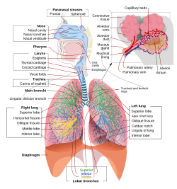
A complete, schematic view of the human respiratory system with their parts and functions.
| |
| Details | |
| Latin | systema respiratorium |
| Identifiers | |
| TA | A06.0.00.000 |
| FMA | 7158 |
| Anatomical terminology | |
The respiratory system (called also respiratory apparatus, ventilatory system) is a biological system consisting of specific organs and structures used for the process of respiration in an organism. The respiratory system is involved in the intake and exchange of oxygen and carbon dioxide between an organism and the environment.
In air-breathing vertebrates like human beings, respiration takes place in the respiratory organs called lungs. The passage of air into the lungs to supply the body with oxygen is known as inhalation, and the passage of air out of the lungs to expel carbon dioxide is known as exhalation; this process is collectively called breathing or ventilation. In humans and other mammals, the anatomical features of the respiratory system include trachea, bronchi, bronchioles, lungs, and diaphragm. Molecules of oxygen and carbon dioxide are passively exchanged, by diffusion, between the gaseous external environment and theblood. This exchange process occurs in the alveoli air sacs in the lungs.[1]
In fish and many invertebrates, respiration takes place through the gills. Other animals, such as insects, have respiratory systems with very simple anatomical features, and in amphibians even the skin plays a vital role in gas exchange.Plants also have respiratory systems but the directionality of gas exchange can be opposite to that in animals. The respiratory system in plants also includes anatomical features such as holes on the undersides of leaves known asstomata.[2]
Contents
[hide]Comparative anatomy and physiology
Horses
Main article: Respiratory system of the horse
Horses are obligate nasal breathers which means that they are different from many other mammals because they do not have the option of breathing through their mouths and must take in oxygen through their noses.
Elephants
The elephant is the only animal known to have no pleural space. Rather, the parietal and visceral pleura are both composed of dense connective tissue and joined to each other via loose connective tissue.[3] This lack of a pleural space, along with an unusually thick diaphragm, are thought to be evolutionary adaptations allowing the elephant to remain underwater for long periods of time while breathing through its trunk which emerges as a snorkel.[4]
Birds
Main article: Bird anatomy § Respiratory system
The respiratory system of birds differs significantly from that found in mammals, containing unique anatomical features such as air sacs. The lungs of birds also do not have the capacity to inflate as birds lack a diaphragm and a pleural cavity. Gas exchange in birds occurs between air capillaries and blood capillaries, rather than in alveoli.
Reptiles
The anatomical structure of the lungs is less complex in reptiles than in mammals, with reptiles lacking the very extensive airway tree structure found in mammalian lungs. Gas exchange in reptiles still occurs in alveoli however, reptiles do not possess a diaphragm. Thus, breathing occurs via a change in the volume of the body cavity which is controlled by contraction of intercostal muscles in all reptiles except turtles. In turtles, contraction of specific pairs of flank muscles governsinspiration or expiration.[5]
Amphibians
Both the lungs and the skin serve as respiratory organs in amphibians. The skin of these animals is highly vascularized and moist, with moisture maintained via secretion of mucus from specialized cells. While the lungs are of primary importance to breathing control, the skin's unique properties aid rapid gas exchange when amphibians are submerged in oxygen-rich water.[6]
Fish
In most fish, respiration takes place through gills. (See also aquatic respiration.) Lungfish, however, do possess one or two lungs. The labyrinth fish have developed a special organ that allows them to take advantage of the oxygen of the air.
Anatomy in invertebrates
Arthropods
Some species of crab use a respiratory organ called a branchiostegal lung.[7] Its gill tissue is formed so as to increase the surface area and the lung is more suited to taking oxygen from the air than from water. Some of the smallest spiders andmites can breathe simply by exchanging gas through the surface of the body. Larger spiders, scorpions and otherarthropods use a primitive book lung.
Insects
Main article: Respiratory system of insects
Most insects breath passively through their spiracles (special openings in the exoskeleton) and the air reaches the body by means of a series of smaller and smaller pipes called 'trachaea' when their diameter is relatively large and 'tracheoles' when their diameter is very small. Diffusion of gases is effective over small distances but not over larger ones, this is one of the reasons insects are all relatively small. Insects which do not have spiracles and trachaea, such as some Collembola, breathe directly through their skins, also by diffusion of gases.[8] The number of spiracles an insect has is variable between species, however they always come in pairs, one on each side of the body, and usually one per segment. Some of the Diplura have eleven, with four pairs on the thorax, but in most of the ancient forms of insects, such as Dragonflies and Grasshoppers there are two thoracic and eight abdominal spiracles. However, in most of the remaining insects there are less. It is at this level of the tracheoles that oxygen is delivered to the cells for respiration. The trachea are water-filled due to the permeable membrane of the surrounding tissues. During exercise, the water level retracts due to the increase in concentration of lactic acid in the muscle cells. This lowers the water potential and the water is drawn back into the cells via osmosis and air is brought closer to the muscle cells. The diffusion pathway is then reduced and gases can be transferred more easily.
Insects were once believed to exchange gases with the environment continuously by the simple diffusion of gases into the tracheal system. More recently, however, large variation in insect ventilatory patterns have been documented and insect respiration appears to be highly variable. Some small insects do demonstrate continuous respiration and may lack muscular control of the spiracles. Others, however, utilize muscular contraction of the abdomen along with coordinated spiracle contraction and relaxation to generate cyclical gas exchange patterns and to reduce water loss into the atmosphere. The most extreme form of these patterns is termed discontinuous gas exchange cycles (DGC).[9]
Molluscs
Molluscs generally possess gills that allow exchange of oxygen from an aqueous environment into the circulatory system. These animals also possess a heart that pumps blood which contains hemocyaninine as its oxygen-capturing molecule. Hence, this respiratory system is similar to that of vertebrate fish. The respiratory system of gastropods can include either gills or a lung.
Physiology in mammals
See also: Respiratory physiology and Respiration (physiology)
Ventilation
In respiratory physiology, ventilation (or ventilation rate) is the rate at which gas enters or leaves the lung. It is categorized under the following definitions:
| Measurement | Equation | Description |
|---|---|---|
| Minute ventilation | tidal volume * respiratory rate[1][2] | the total volume of gas entering the lungs per minute. |
| Alveolar ventilation | (tidal volume – dead space) * respiratory rate [1] | the volume of gas per unit time that reaches the alveoli, the respiratory portions of the lungs where gas exchange occurs. |
| Dead space ventilation | dead space * respiratory rate[3] | the volume of gas per unit time that does not reach these respiratory portions, but instead remains in the airways (trachea, bronchi, etc.). |
Control
Ventilation occurs under the control of the autonomic nervous system from parts of the brain stem, the medulla oblongataand the pons. This area of the brain forms the respiration regulatory center, a series of interconnected brain cells within the lower and middle brain stem which coordinate respiratory movements. The sections are the pneumotaxic center, theapneustic center, and the dorsal and ventral respiratory groups. This section is especially sensitive during infancy, and the neurons can be destroyed if the infant is dropped and/or shaken violently. The result can be death due to "shaken baby syndrome".[10]
The breathing rate increases with the concentration of carbon dioxide in the blood, which is detected by peripheralchemoreceptors in the aorta and carotid artery and central chemoreceptors in the medulla. Exercise also increases respiratory rate, due to the action of proprioceptors, the increase in body temperature, the release of epinephrine, and motor impulses originating from the brain.[11] In addition, it can increase due to increased inflation in the lungs, which is detected by stretch receptors.
Inhalation
Inhalation is initiated by the diaphragm and supported by the external intercostal muscles. Normal resting respirations are 10 to 18 breaths per minute, with a time period of 2 seconds. During vigorous inhalation (at rates exceeding 35 breaths per minute), or in approaching respiratory failure, accessory muscles of respiration are recruited for support. These consist ofsternocleidomastoid, platysma, and the scalene muscles of the neck. Pectoral muscles and latissimus dorsi are also accessory muscles.
Under normal conditions, the diaphragm is the primary driver of inhalation. When the diaphragm contracts, the ribcageexpands and the contents of the abdomen are moved downward. This results in a larger thoracic volume and negative pressure (with respect to atmospheric pressure) inside the thorax. As the pressure in the chest falls, air moves into the conducting zone. Here, the air is filtered, warmed, and humidified as it flows to the lungs.
During forced inhalation, as when taking a deep breath, the external intercostal muscles and accessory muscles aid in further expanding the thoracic cavity. During inhalation the diaphragm contracts.
Exhalation
Exhalation is generally a passive process; however, active or forced exhalation is achieved by the abdominal and theinternal intercostal muscles. During this process air is forced or exhaled out.
The lungs have a natural elasticity: as they recoil from the stretch of inhalation, air flows back out until the pressures in the chest and the atmosphere reach equilibrium.[12]
During forced exhalation, as when blowing out a candle, expiratory muscles including the abdominal muscles and internal intercostal muscles, generate abdominal and thoracic pressure, which forces air out of the lungs.
Gas exchange
The major function of the respiratory system is gas exchange between the external environment and an organism'scirculatory system. In humans and other mammals, this exchange facilitates oxygenation of the blood with a concomitant removal of carbon dioxide and other gaseous metabolic wastes from the circulation.[13] As gas exchange occurs, the acid-base balance of the body is maintained as part of homeostasis. If proper ventilation is not maintained, two opposing conditions could occur: respiratory acidosis, a life-threatening condition, and respiratory alkalosis.
Upon inhalation, gas exchange occurs at the alveoli, the tiny sacs which are the basic functional component of the lungs. The alveolar walls are extremely thin (approx. 0.2 micrometres). These walls are composed of a single layer of epithelial cells (type I and type II epithelial cells) close to the pulmonary capillaries which are composed of a single layer of endothelial cells. The close proximity of these two cell types allows permeability to gases and, hence, gas exchange. This whole mechanism of gas exchange is carried by the simple phenomenon of pressure difference. When the air pressure is high inside the lungs, the air from lungs flow out. When the air pressure is low inside, then air flows into the lungs.
Immune functions
Airway epithelial cells can secrete a variety of molecules that aid in the defense of lungs. Secretory immunoglobulins (IgA), collectins (including Surfactant A and D), defensins and other peptides and proteases, reactive oxygen species, and reactive nitrogen species are all generated by airway epithelial cells. These secretions can act directly as antimicrobials to help keep the airway free of infection. Airway epithelial cells also secrete a variety of chemokines and cytokines that recruit the traditional immune cells and others to site of infections.
Most of the respiratory system is lined with mucous membranes that contain mucosal-associated lymphoid tissue, which produces white blood cells such as lymphocytes.
Metabolic and endocrine functions of the lungs
In addition to their functions in gas exchange, the lungs have a number of metabolic functions. They manufacture surfactant for local use, as noted above. They also contain a fibrinolytic system that lyses clots in the pulmonary vessels. They release a variety of substances that enter the systemic arterial blood and they remove other substances from the systemic venous blood that reach them via the pulmonary artery. Prostaglandins are removed from the circulation, but they are also synthesized in the lungs and released into the blood when lung tissue is stretched. The lungs also activate one hormone; the physiologically inactive decapeptide angiotensin I is converted to the pressor, aldosterone-stimulating octapeptide angiotensin II in the pulmonary circulation. The reaction occurs in other tissues as well, but it is particularly prominent in the lungs. Large amounts of the angiotensin-converting enzyme responsible for this activation are located on the surface of the endothelial cells of the pulmonary capillaries. The converting enzyme also inactivates bradykinin. Circulation time through the pulmonary capillaries is less than one second, yet 70% of the angiotensin I reaching the lungs is converted to angiotensin II in a single trip through the capillaries. Four other peptidases have been identified on the surface of the pulmonary endothelial cells.
Vocalization
The movement of gas through the larynx, pharynx and mouth allows humans to speak, or phonate. Vocalization, or singing, in birds occurs via the syrinx, an organ located at the base of the trachea. The vibration of air flowing across the larynx (vocal cords), in humans, and the syrinx, in birds, results in sound. Because of this, gas movement is extremely vital forcommunication purposes.
Temperature control
Panting in dogs, cats and some other animals provides a means of controlling body temperature. This physiological response is used as a cooling mechanism.
Coughing and sneezing
Irritation of nerves within the nasal passages or airways, can induce a cough reflex and sneezing. These responses cause air to be expelled forcefully from the trachea or nose, respectively. In this manner, irritants caught in the mucus which lines the respiratory tract are expelled or moved to the mouth where they can be swallowed. During coughing, contraction of the smooth muscle narrows the trachea by pulling the ends of the cartilage plates together and by pushing soft tissue out into the lumen. This increases the expired airflow rate to dislodge and remove any irritant particle or mucus.
Development
Humans and mammals
The respiratory system lies dormant in the human fetus during pregnancy. At birth, the respiratory system becomes fully functional upon exposure to air, although some lung development and growth continues throughout childhood.[14] Pre-term birth can lead to infants with under-developed lungs. These lungs show incomplete development of the alveolar type II cells, cells that produce surfactant. The lungs of pre-term infants may not function well because the lack of surfactant leads to increased surface tension within the alveoli. Thus, many alveoli collapse such that no gas exchange can occur within some or most regions of an infant's lungs, a condition termed respiratory distress syndrome. Basic scientific experiments, carried out using cells from chicken lungs, support the potential for using steroids as a means of furthering development of type II alveolar cells.[15] In fact, once a pre-mature birth is threatened, every effort is made to delay the birth, and a series of steroidshots is frequently administered to the mother during this delay in an effort to promote lung growth.[16]
Disease
Disorders of the respiratory system can be classified into four general areas:
- Obstructive conditions (e.g., emphysema, bronchitis, asthma)
- Restrictive conditions (e.g., fibrosis, sarcoidosis, alveolar damage, pleural effusion)
- Vascular diseases (e.g., pulmonary edema, pulmonary embolism, pulmonary hypertension)
- Infectious, environmental and other "diseases" (e.g., pneumonia, tuberculosis, asbestosis, particulate pollutants):
Coughing is of major importance, as it is the body's main method to remove dust, mucus, saliva, and other debris from the lungs. Inability to cough can lead to infection. Deep breathing exercises may help keep finer structures of the lungs clear from particulate matter, etc.
The respiratory tract is constantly exposed to microbes due to the extensive surface area, which is why the respiratory system includes many mechanisms to defend itself and prevent pathogens from entering the body.
Disorders of the respiratory system are usually treated internally by a pulmonologist and Respiratory Therapist.
Plants
Plants use carbon dioxide gas in the process of photosynthesis, and exhale oxygen gas as waste. The chemical equation of photosynthesis is 6 CO2 (carbon dioxide) and 6 H2O (water) and that makes 6 O2 (oxygen) and C6H12O6 (glucose). What is not expressed in the chemical equation is the capture of energy from sunlight which occurs. Photosynthesis uses electrons on the carbon atoms as the repository for that energy. Respiration is the opposite of photosynthesis. It reclaims the energy to power chemical reactions in cells. In so doing the carbon atoms and their electrons are combined with oxygen forming a gas which is easily removed from both the cells and the organism. Plants use both processes, photosynthesis to capture the energy and respiration to use it.
Plant respiration is limited by the process of diffusion. Plants take in carbon dioxide through holes on the undersides of theirleaves known as stoma or pores. However, most plants require little air.[citation needed] Most plants have relatively few living cells outside of their surface because air (which is required for metabolic content) can penetrate only skin deep. However, most plants are not involved in highly aerobic activities, and thus have no need of these living cells.
Lung
From Wikipedia, the free encyclopedia
For other uses, see Lung (disambiguation).
This article is about lungs in general. For human lungs, see Human lung.
"Lunged" redirects here. For other uses, see Lunge.
The lung is the essential respiration organ in many air-breathing animals, including most tetrapods, a few fish and a few snails. In mammals and the more complex life forms, the two lungs are located near the backbone on either side of the heart. Their principal function is to transport oxygen from the atmosphere into the bloodstream, and to release carbon dioxide from the bloodstream into the atmosphere. A large surface area is needed for this exchange of gases which is accomplished by the mosaic of specialized cells that form millions of tiny, exceptionally thin-walled air sacs called alveoli.
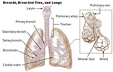
To understand the anatomy of the lungs, the passage of air through the nose and mouth to the alveoli must be studied. The progression of air through either the mouth or the nose, travels through the nasopharynx and oropharynx of the pharynx,larynx, and the trachea (windpipe). The air passes down the trachea, which divides into two main bronchi; these branch to the left and right lungs where they progressively subdivide into a system of bronchi and bronchioles until the alveoli are reached. These many alveoli are where the gas exchange of carbon dioxide and oxygen takes place.[2]
Breathing is driven by muscular action; in early tetrapods, air was driven into the lungs by the pharyngeal muscles via buccal pumping, which is still found in amphibians. Reptiles, birds and mammals use their musculoskeletal system to support and foster breathing.
Medical terms related to the lung often begin with pulmo-, such as in the (adjectival form: pulmonary) or from the Latin pulmonarius ("of the lungs"), or with pneumo-(from Greek πνεύμων "lung").
Contents
[hide]Mammalian lungs
See also: Human lung
The lungs of mammals including those of humans, have a soft, spongelike texture and are honeycombed with epithelium, having a much larger surface area in total than the outer surface area of the lung itself.
Breathing is largely driven by the muscular diaphragm at the bottom of the thorax. Contraction of the diaphragm pulls the bottom of the cavity in which the lung is enclosed downward, increasing volume and thus decreasing pressure, causing air to flow into the airways. Air enters through the oral and nasal cavities; it flows through the pharynx, then the larynx and into the trachea, which branches out into the main bronchi and then subsequent divisions. During normalbreathing, expiration is passive and no muscles are contracted (the diaphragm relaxes). The rib cage itself is also able to expand and contract to some degree through the use of the intercostal muscles, together with the action of other respiratory and accessory respiratory muscles. As a result, air is transported into or expelled out of the lungs. This type of lung is known as a bellows lung as it resembles a blacksmith's bellows.[3]
Anatomy
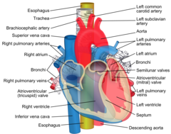
In humans, the trachea divides into the two main bronchi that enter the roots of the lungs. The bronchi continue to divide within the lung, and after multiple divisions, give rise to bronchioles. The bronchial tree continues branching until it reaches the level of terminal bronchioles, which lead to alveolar sacs. Alveolar sacs, are made up of clusters of alveoli, like individual grapes within a bunch. The individual alveoli are tightly wrapped in blood vessels and it is here that gas exchange actually occurs. Deoxygenated blood from the heart is pumped through the pulmonary artery to the lungs, where oxygen diffusesinto blood and is exchanged for carbon dioxide in the haemoglobin of the erythrocytes. The oxygen-rich blood returns to the heart via the pulmonary veins to be pumped back into systemic circulation."Lung Disease & Respiratory Health Center".
Human lungs are located in two cavities on either side of the heart. Though similar in appearance, the two are not identical. Both are separated into lobes by fissures, with three lobes on the right and two on the left. The lobes are further divided into segments and then into lobules, hexagonal divisions of the lungs that are the smallest subdivision visible to the naked eye.[citation needed] The connective tissue that divides lobules is often blackened in smokers. The medial border of the right lung is nearly vertical, while the left lung contains a cardiac notch. The cardiac notch is a concave impression molded to accommodate the shape of the heart.
Each lobe is surrounded by a pleural cavity, which consists of two pleurae. The parietal pleura lies against the rib cage, and the visceral pleura lies on the surface of the lungs. In between the pleura is pleural fluid. The pleural cavity helps to lubricate the lungs, as well as providing surface tension to keep the lung surface in contact with the rib cage.
Lungs are to a certain extent "overbuilt" and have a tremendous reserve volume as compared to the oxygen exchange requirements when at rest. Such excess capacity is one of the reasons that individuals can smoke for years without having a noticeable decrease in lung function while still or moving slowly; in situations like these only a small portion of the lungs are actually perfused with blood for gas exchange. Destruction of too many alveoli over time leads to the condition emphysema, which is associated with extreme shortness of breath. As oxygen requirements increase due to exercise, a greater volume of the lungs is perfused, allowing the body to match its CO2/O2 exchange requirements. Additionally, due to the excess capacity, it is possible for humans to live with only one lung, with the one compensating for the other's loss.
The environment of the lung is very moist, which makes it hospitable for bacteria. Many respiratory illnesses are the result of bacterial or viral infection of the lungs. Inflammation of the lungs is known as pneumonia; inflammation of the pleurasurrounding the lungs is known as pleurisy.
Vital capacity is the maximum volume of air that a person can exhale after maximum inhalation; it can be measured with aspirometer. In combination with other physiological measurements, the vital capacity can help make a diagnosis of underlying lung disease.
The lung parenchyma is strictly used to refer solely to alveolar tissue with respiratory bronchioles, alveolar ducts andterminal bronchioles.[4] However, it often includes any form of lung tissue, also including bronchioles, bronchi, blood vesselsand lung interstitium.[4]
Non respiratory functions
In addition to their function in respiration, the lungs also:
- Alter the pH of blood by facilitating alterations in the partial pressure of carbon dioxide
- Filter out small blood clots formed in veins
- Filter out gas micro-bubbles occurring in the venous blood stream such as those created during decompression afterunderwater diving.[5]
- Influence the concentration of some biologic substances and drugs used in medicine in blood
- Convert angiotensin I to angiotensin II by the action of angiotensin-converting enzyme
- May serve as a layer of soft, shock-absorbent protection for the heart, which the lungs flank and nearly enclose.
- Immunoglobulin-A is secreted in the bronchial secretion and protects against respiratory infections.
- Maintain sterility by producing mucus containing antimicrobial compounds.[6] Mucus contains glycoproteins, e.g., mucins,lactoferrin,[7] lysozyme, lactoperoxidase.[8][9] We find also on the epithelium Dual oxidase 2[10][11][12] proteins generating hydrogen peroxide, useful for hypothiocyanite endogenous antimicrobial synthesis. Function not in place in cystic fibrosis patient lungs.[13][14]
- Ciliary escalator action is an important defence system against air-borne infection. The dust particles and bacteria in the inhaled air are caught in the mucous layer present at the mucosal surface of respiratory passages and are moved up towards pharynx by the rhythmic upward beating action of the cilia.
- Provide airflow for the creation of vocal sounds.
- The lungs serve as a reservoir of blood in the body. The blood volume of the lungs is about 450 milliliters on average, about 9 percent of the total blood volume of the entire circulatory system. This quantity can easily fluctuate from between one-half and twice the normal volume. Loss of blood from the systemic circulation by hemorrhage can be partially compensated for by shunting blood from the lungs into the systemic vessels[15]
- Thermoregulation via panting (observed in some animals, but not humans)
Avian lungs
Avian lungs do not have alveoli as mammalian lungs do; birds have honey-comb-like, faveolar lungs[citation needed], which contain millions of tiny passages called parabronchi. There are air vesicles, called atria, which project radially from the walls of the parabronchi. The gas exchange tissues are set into the walls of the atria and gases travel via diffusion between the gas exchange tissues and the lumen of each parabronchus. There are two categories of parabronchi. The paleopulmonic parabronchi are found in all birds and air flows through them in the same direction—posterior to anterior during inhalation and exhalation. Some bird species also have neopulmonic parabronchi where the air flow is bidirectional. The paleopulmonic unidirectional airflow is in contrast to the mammalian system, in which the direction of airflow in the lung is tidal, reversing between inhalation and exhalation.
By utilizing a unidirectional flow of air, avian lungs are able to extract a greater concentration of oxygen from inhaled air. Birds are thus equipped to fly at altitudes at which mammals would succumb to hypoxia. This also allows them to sustain a higher metabolic rate than most equivalent weight mammals.[16] Note that some species of small bats have a higher mean total morphometric pulmonary diffusing capacity for oxygen than equivalent weight birds but this is the exception and is not the rule.[17]
The lungs of birds are relatively small, but are connected to 8–9 air sacs that extend through much of the body, and are in turn connected to air spaces within the bones. The air sacs, although thin walled, are poorly vascularized, and do not contribute much to gas exchange, but they do act like bellows to ventilate the lungs. The air sacs expand and contract due to changes in the volume of the combined thorax and abdominal cavity. This volume change is caused by the movement of the sternum and ribs and this movement is often synchronized with movement of the flight muscles.[18]
Because of the complexity of the system, misunderstanding is common and it is incorrectly believed that it takes two breathing cycles for air to pass entirely through a bird's respiratory system. Air is not stored in either the posterior or anterior sacs between respiration cycles, air moves continuously from the posterior to the anterior of the lungs throughout respiration. This type of lung construction is called a circulatory lung, as distinct from the bellows lung possessed by other animals.[16]
Reptilian lungs
Main article: Reptile anatomy § Respiration
Reptilian lungs are typically ventilated by a combination of expansion and contraction of the ribs via axial muscles and buccal pumping. Crocodilians also rely on the hepatic piston method, in which the liver is pulled back by a muscle anchored to the pubic bone (part of the pelvis), which in turn pulls the bottom of the lungs backward, expanding them. Turtles, which are unable to move their ribs, instead use their forelimbs and pectoral girdle to force air in and out of the lungs.[18]
The lung of most reptiles has a single bronchus running down the centre, from which numerous branches reach out to individual pockets throughout the lungs. These pockets are similar to, but much larger and fewer in number than, mammalian alveoli, and give the lung a sponge-like texture. In tuataras, snakes, and some lizards, the lungs are simpler in structure, similar to that of typical amphibians.[18]
Snakes and limbless lizards typically possess only the right lung as a major respiratory organ; the left lung is greatly reduced, or even absent. Amphisbaenians, however, have the opposite arrangement, with a major left lung, and a reduced or absent right lung.[18]
Both crocodilians and monitor lizards have developed lungs similar to those of birds, providing an unidirectional airflow and even possessing air sacs.[19] The now extinct pterosaurs have seemingly even further refined this type of lung, extending the airsacs into the wing membranes and, in the case of Pteranodontia, the hindlimbs.
Amphibian lungs
The lungs of most frogs and other amphibians are simple balloon-like structures, with gas exchange limited to the outer surface area of the lung. This is not a very efficient arrangement, but amphibians have low metabolic demands and can also quickly dispose of carbon dioxide by diffusion across their skin in water, and supplement their oxygen supply by the same method. Unlike higher vertebrates, who use a breathing system driven by negative pressure where the lungs are inflated by expanding the rib cage, amphibians employ positive pressure system, forcing air down into the lungs by buccal pumping.[20]The floor of the mouth is lowered, filling the mouth cavity with air. The throat muscles then presses the throat against the underside of the skull, forcing the air into the lungs.[21]
Due to the possibility of respiration across the skin combined with small size, all known lungless tetrapods are amphibians. The majority of salamander species are lungless salamanders, which respirate through their skin and tissues lining their mouth. This necessarily restrict their size, all are small and rather thread-like in appearance, maximizing skin surface relative to body volume.[22] The only other known lungless tetrapods are the Bornean Flat-headed Frog (Barbourula kalimantanensis) and Atretochoana eiselti, a caecilian.
The lungs of amphibians typically have a few narrow septa of soft tissue around the outer walls, increasing the respiratory surface area and giving the lung a honey-comb appearance. In some salamanders even these are lacking, and the lung has a smooth wall. In caecilians, as in snakes, only the right lung attains any size or development.[18]
Lungfish
The lungs of lungfish are similar to those of amphibians, with few, if any, internal septa. In the Australian lungfish, there is only a single lung, albeit divided into two lobes. Other lungfish and Polypterus, however, have two lungs, which are located in the upper part of the body, with the connecting duct curving round and above the esophagus. The blood supply also twists around the esophagus, suggesting that the lungs originally evolved in the ventral part of the body, as in other vertebrates.[18]
Invertebrate lungs
Some invertebrates have "lungs" that serve a similar respiratory purpose as, but are not evolutionarily related to, vertebrate lungs. Some arachnids have structures called "book lungs" used for atmospheric gas exchange. The Coconut crab uses structures called Branchiostegal lungs to breathe air and indeed will drown in water, hence it breathes on land and holds its breath underwater. The Pulmonata are an order of snails and slugs that have developed "lungs".
Origins of the vertebrate lung
The lungs of today's terrestrial vertebrates and the gas bladders of today's fish are believed to have evolved from simple sacs (outpocketings) of the esophagus that allowed early fish to gulp air under oxygen-poor conditions.[23] These outpocketings first arose in the bony fish. In most of the ray-finned fish the sacs evolved into closed off gas bladders, while a number of carps, trouts, herrings, catfish, eels has retained the physostome condition with the sack being open to the esophagus. In more basal bony fish, such as the gar, bichir, bowfin and the lobe-finned fish, the bladders have evolved to primarily function as lungs.[23] The lobe-finned fish gave rise to the land-based tetrapods. Thus, the lungs of vertebrates arehomologous to the gas bladders of fish (but not to their gills). This is reflected by the fact that the lungs of a fetus also develop from an outpocketing of the esophagus and in the case of the physostome gas bladders, which can serve as bothbuoyancy organ and with the pneumatic duct to the gut also serve as lungs. This condition is found in more "primitive"teleosts, and is lost in the higher orders. (This is an instance of correlation between ontogeny and phylogeny.) No known animals have both a gas bladder and lungs.
Trachea
For the genus of moth, see Trachea (moth).
| Trachea | |
|---|---|
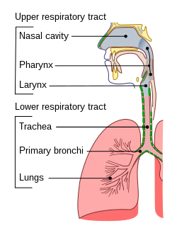
Conducting passages.
| |
| Details | |
| tracheal branches of inferior thyroid artery | |
| brachiocephalic vein, azygos veinaccessory hemiazygos vein | |
| Identifiers | |
| Anatomical terminology | |
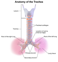
The trachea, colloquially called windpipe, is a tube that connects the pharynxand larynx to the lungs, allowing the passage of air, and so is present in all air-breathing animals with lungs. Only in the lungfish, where the lung is connected to the pharynx and the larynx, it is absent.[citation needed] The trachea extends from the larynx and branches into the two primary bronchi. At the top of the trachea the cricoid cartilage attaches it to the larynx. This is the only complete ring, the others being incomplete rings of reinforcing cartilage. The trachealis muscle joins the ends of the rings and these are joined vertically by bands of fibrous connective tissue, the annular ligaments of trachea. The epiglottiscloses the opening to the larynx during swallowing.
The trachea develops in the second month of development. It is lined with anepithelium that has goblet cells which produce protective mucins. Aninflammatory condition, also involving the larynx and bronchi, called croup can result in a barking cough. A tracheotomy is often performed for ventilation in surgical operations where needed. Intubation is also carried out for the same reason by the inserting of a tube into the trachea. From 2008, successful operations have transplanted a windpipe grown by stem cells, and synthetic windpipes.
Contents
[hide]Structure
The human trachea has an inner diameter of about 25 millimetres (1 in) and a length of about 10 to 16 centimetres (4 to 6 in). It commences at the lower border of the larynx, level with the sixth cervical vertebra, and bifurcates into the primary bronchi at the vertebral level of thoracic vertebraT5, or up to two vertebrae lower or higher, depending on breathing. A ring of hyaline cartilage called thecricoid cartilage forms the inferior wall of the larynx and is attached to the top of trachea. The cricoid cartilage is the only complete ring of cartilage in the trachea.
There are from fifteen to twenty incomplete C-shaped tracheal rings of cartilage that reinforce the front and sides of the trachea to protect and maintain the airway. This leaves a membranous wall (pars membranacea) at the back, without cartilage. The trachealis muscle connects the ends of the incomplete rings and contracts during coughing, reducing the size of the lumen of the trachea to increase the rate of air flow.[1] The esophagus lies posteriorly to the trachea. Circular bands of fibrous connective tissue called the annular ligaments of trachea join the tracheal rings together. The cartilaginous rings are incomplete to allow the trachea to collapse slightly so that food can pass down the esophagus. A flap-like epiglottiscloses the opening to the larynx during swallowing to prevent swallowed matter from entering the trachea.
Variations
Tracheal agenesis,[2] is a rare birth defect in which the trachea fails to develop. The defect is usually fatal though sometimes surgical intervention has been successful.
A tracheoesophageal fistula is a congenital defect in which the trachea and esophagus are abnormally connected.
Development
In the fourth week of embryogenesis as the respiratory bud grows, the trachea separates from the foregut through the formation of tracheoesophageal ridges which fuse to form the tracheoesophageal septum and this separates the future trachea from the oesophagus and divides the foregut tube into the laryngotracheal tube. Before the end of the fifth week, the trachea begins to develop from the laryngotracheal tube which develops from the laryngotracheal groove. The first part of the cephalic region of the tube forms the larynx, and the next part forms the trachea.
Histology[edit]
The trachea is lined with pseudostratified columnar epithelium a type of epitheliumwith goblet cells that produce mucins the main component of mucus, to moisten and protect the airways.[3] Mucus lines the ciliated cells of the trachea to trap inhaled foreign particles that the cilia then waft upward toward the larynx and then the pharynx where it can be either swallowed into the stomach or expelled as phlegm.
Clinical significance
Inflammation
Inflammation of the trachea is known as tracheitis. When the trachea is inflamed as well as the larynx and bronchi, this is known as croup, which often causes a distinct, barking cough.
Intubation
Tracheal intubation refers to the insertion of a tracheal tube down the trachea. This procedure is commonly performed during surgery, in order to ensure a person receives enough oxygen when sedated. The tube inserted down the trachea is connected to a machine that monitors the airflow, oxygenation and several other metrics. This is often one of the responsibilities of an anesthetist during surgery.
In an emergency, or when tracheal intubation is deemed impossible, a tracheotomy is often performed to insert a tube for ventilation, usually when needed for particular types of surgery to be carried out so that the airway is kept open for sufficient time. Another less invasive method is used when a procedure can be carried out more quickly, or in an emergency situation, and this is a cricothyrotomy.
Other[edit]
- Asphyxia
- Tracheomalacia (weakening of the tracheal cartilage)
- Tracheal collapse (in dogs)
- Tracheobronchial injury (perforation of the trachea or bronchi)
- Mounier-Kuhn syndrome (an abnormal enlargement of the trachea)
Other animals[edit]
Allowing for variations in the length of the neck, the trachea in other mammals is, in general, similar to that in humans. Generally, it is also similar to the reptilian trachea.[4]
Vertebrates
In birds, the trachea runs from the pharynx to the syrinx, from which the primary bronchi diverge. Swans have an unusually elongated trachea, part of which is coiled beneath the sternum; this may act as a resonator to amplify sound. In some birds, the tracheal rings are complete, and may even be ossified.[4]
In amphibians, the trachea is normally extremely short, and leads directly into the lungs, without clear primary bronchi. A longer trachea is, however, found in some long-necked salamanders, and in caecilians. While there are irregular cartilagenous nodules on the amphibian trachea, these do not form the rings found in amniotes.[4]
The only vertebrate to have lungs, but no trachea, is Polypterus, in which the lungs arise directly from the pharynx.[4]
Invertebrates
The invertebrate trachea refers to the open respiratory system composed ofspiracles, tracheae, and tracheoles that terrestrial arthropods have to transportmetabolic gases to and from tissues.[5] The distribution of spiracles can vary greatly among the many orders of insects, but in general each segment of the body can have only one pair of spiracles, each of which connects to an atrium and has a relatively large tracheal tube behind it. The tracheae are invaginations of the cuticular exoskeleton that branch (anastomose) throughout the body with diameters from only a few micrometres up to 0.8 mm. The smallest tubes, tracheoles, penetrate cells and serve as sites of diffusion for water, oxygen, and carbon dioxide. Gas may be conducted through the respiratory system by means of active ventilation or passive diffusion. Unlike vertebrates, insects do not generally carry oxygen in theirhaemolymph.[6] This is one of the factors that may limit their size.
A tracheal tube may contain ridge-like circumferential rings of taenidia in variousgeometries such as loops or helices. In the head, thorax, or abdomen, tracheae may also be connected to air sacs. Many insects, such as grasshoppers and bees, which actively pump the air sacs in their abdomen, are able to control the flow of air through their body. In some aquatic insects, the tracheae exchange gas through the body wall directly, in the form of a gill, or function essentially as normal, via aplastron. Note that despite being internal, the tracheae of arthropods are shed during moulting (ecdysis).[citation needed]
History[edit]
In 2008, a Colombian woman received a trachea transplant using her own stem cells so her body would not reject the transplant.[7] In June 2011, a team of surgeons led by Professor Paolo Macchiarini at the Karolinska University Hospital performed the first synthetic windpipe transplant on a 36-year-old Eritrean man, Andemariam Teklesenbet Beyene.[8][9]












No comments:
Post a Comment